Σφακιανάκης Αλέξανδρος
ΩτοΡινοΛαρυγγολόγος
Αναπαύσεως 5 Άγιος Νικόλαος
Κρήτη 72100
00302841026182
00306932607174
alsfakia@gmail.com

Αρχειοθήκη ιστολογίου

! # Ola via Alexandros G.Sfakianakis on Inoreader

Η λίστα ιστολογίων μου

Πέμπτη 23 Μαΐου 2019

Severe abdominal pain out of proportion to physical examination findings should be assumed to be AMI until disproven.Clinical scenario differentiates AMI as mesenteric arterial emboli, mesenteric arterial thrombosis, NOMI, or mesenteric venous thrombosis.

Acute mesenteric ischemia: guidelines of the World Society of Emergency Surgery

  • Miklosh BalaEmail author,
  • Jeffry Kashuk,
  • Ernest E. Moore,
  • Yoram Kluger,
  • Walter Biffl,
  • Carlos Augusto Gomes,
  • Offir Ben-Ishay,
  • Chen Rubinstein,
  • Zsolt J. Balogh,
  • Ian Civil,
  • Federico Coccolini,
  • Ari Leppaniemi,
  • Andrew Peitzman,
  • Luca Ansaloni,
  • Michael Sugrue,
  • Massimo Sartelli,
  • Salomone Di Saverio,
  • Gustavo P. Fraga and
  • Fausto Catena
World Journal of Emergency Surgery201712:38

https://doi.org/10.1186/s13017-017-0150-5

  • Received: 30 June 2017
  • Accepted: 1 August 2017
  • Published: 7 August 2017

Abstract

Acute mesenteric ischemia (AMI) is typically defined as a group of diseases characterized by an interruption of the blood supply to varying portions of the small intestine, leading to ischemia and secondary inflammatory changes. If untreated, this process will eventuate in life threatening intestinal necrosis. The incidence is low, estimated at 0.09–0.2% of all acute surgical admissions. Therefore, although the entity is an uncommon cause of abdominal pain, diligence is always required because if untreated, mortality has consistently been reported in the range of 50%. Early diagnosis and timely surgical intervention are the cornerstones of modern treatment and are essential to reduce the high mortality associated with this entity. The advent of endovascular approaches in parallel with modern imaging techniques may provide new options. Thus, we believe that a current position paper from World Society of Emergency Surgery (WSES) is warranted, in order to put forth the most recent and practical recommendations for diagnosis and treatment of AMI. This review will address the concepts of AMI with the aim of focusing on specific areas where early diagnosis and management hold the strongest potential for improving outcomes in this disease process.

Some of the key points include the prompt use of CT angiography to establish the diagnosis, evaluation of the potential for revascularization to re-establish blood flow to ischemic bowel, resection of necrotic intestine, and use of damage control techniques when appropriate to allow for re-assessment of bowel viability prior to definitive anastomosis and abdominal closure.

Keywords

  • Mesenteric ischemia
  • Mesenteric arterial occlusion
  • Mesenteric angiography
  • Mesenteric artery stenting
  • Small bowel ischemia
  • Guidelines
  • Recommendations

Background

Acute mesenteric ischemia (AMI) may be defined as a sudden interruption of the blood supply to a segment of the small intestine, leading to ischemia, cellular damage, intestinal necrosis, and eventually patient death if untreated [1]. AMI may be non-occlusive (NOMI) or occlusive, with the primary etiology further defined as mesenteric arterial embolism (50%), mesenteric arterial thrombosis (15–25%), or mesenteric venous thrombosis (5–15%) [2, 3]. The overall incidence is low (0.09 to 0.2% of all acute admissions to emergency departments), representing an uncommon cause of abdominal pain [4, 5, 6]. Prompt diagnostic and intervention are essential to reduce the high mortality rates (50 to 80%) [7, 8, 9, 10].

There are currently no level 1 evidence to guide the evaluation and treatment of suspected AMI, and the published literature contains primarily institutional reviews, case series and personal recommendations with no clearly defined treatment guidelines.

Accordingly, this review aims to provide an update with recommendations based on the most currently accepted concepts in the management of AMI.

The current presentation evolved from the contributions of a group of experts in the field who submitted their evidence-based literature review of key points pertaining to diagnosis and management of AMI. Following preliminary preparation of these key points, a coordinated presentation was organized during the WSES World Congress, May 2017 in Campinas, Brazil. The final version has taken into account the presentations at the congress as well as pertinent group discussions and comments on the various presentations.

The grading of recommendations was evaluated (Table 1).
Table 1

Grading of recommendations

Grade of recommendation

Clarity of risk/benefit

Quality of supporting evidence

Implications

1A


Strong recommendation, high-quality evidence

Benefits clearly outweigh risk and burdens, or vice versa

RCTs without important limitations or overwhelming evidence from observational studies

Strong recommendation, applies to most patients in most circumstances without reservation

1B


Strong recommendation, moderate-quality evidence

Benefits clearly outweigh risk and burdens, or vice versa

RCTs with important limitations (inconsistent results, methodological flaws, indirect analyses, or imprecise conclusions) or exceptionally strong evidence from observational studies

Strong recommendation, applies to most patients in most circumstances without reservation

1C


Strong recommendation, low-quality or very low-quality evidence

Benefits clearly outweigh risk and burdens, or vice versa

Observational studies or case series

Strong recommendation but subject to change when higher quality evidence becomes available

2A


Weak recommendation, high-quality evidence

Benefits closely balanced with risks and burden

RCTs without important limitations or overwhelming evidence from observational studies

Weak recommendation, best action may differ depending on the patient, treatment circumstances, or social values

2B


Weak recommendation, moderate-quality evidence

Benefits closely balanced with risks and burden

RCTs with important limitations (inconsistent results, methodological flaws, indirect or imprecise) or exceptionally strong evidence from observational studies

Weak recommendation, best action may differ depending on the patient, treatment circumstances, or social values

2C


Weak recommendation, low-quality or very low-quality evidence

Uncertainty in the estimates of benefits, risks, and burden; benefits, risk, and burden may be closely balanced

Observational studies or case series

Very weak recommendation; alternative treatments may be equally reasonable and merit consideration

Mesenteric vascular anatomy and physiology

The superior mesenteric artery (SMA) is the primary blood supply for the small bowel with some collateral flow from the celiac arterial system, via the superior and inferior pancreaticoduodenal arteries, as well as from the inferior mesenteric artery. Intestinal blood returns via the portal vein. The splanchnic circulation receives 15–35% of the cardiac output, depending on the feeding state, but oxygen extraction is relatively low, accounting for the oxygen delivery capacity of the portal vein to the liver. Thus, blood supply must be reduced by more than 50% before the small intestine becomes ischemic [11].

Furthermore, the intestines can autoregulate oxygen availability via enhanced oxygen extraction and perfusion due to vasodilation. Experimentally, it had been shown that mesenteric ischemia does not occur until the patient's mean arterial pressure is <45 mmHg [12]. Consequently, the small intestine is able to compensate for a 75% reduction in mesenteric blood flow for up to 12 h [13].

Pathophysiology and epidemiology

Acute mesenteric arterial embolism

Roughly, 50% of all cases of AMI are due to acute mesenteric embolism [2, 3]. Mesenteric emboli can originate from the left atrium, associated with cardiac dysrhythmias such as atrial fibrillation, left ventricle with global myocardial dysfunction associated with poor ejection fraction, or cardiac valves due to endocarditis. Occasionally emboli generated from an atherosclerotic aorta. Emboli typically lodge at points of normal anatomic narrowing, and the SMA is particularly vulnerable because of its relatively large diameter and low takeoff angle from the aorta. The majority of emboli lodge 3 to 10 cm distal to the origin of the SMA, thus classically sparing the proximal jejunum and colon. More than 20% of emboli to the SMA are associated with concurrent emboli to another arterial bed including the spleen, or kidney. Thus, findings of changes in these organs on CTA suggest a proximal embolic source [14].

Acute mesenteric arterial thrombosis

Thrombosis of the SMA (approximately 25% of cases) is usually associated with pre-existing chronic atherosclerotic disease leading to stenosis. Many of these patients have a history consistent with chronic mesenteric ischemia (CMI), including postprandial pain, weight loss, or "food fear", and thus a systematic history is important when evaluating a patient suspected to have AMI. Thrombosis usually occurs at the origin of visceral arteries, moreover, an underlying plaque in the SMA usually progresses to a critical stenosis over years resulting in collateral beds. Accordingly, symptomatic SMA thrombosis most often accompanies celiac occlusion [15]. SMA thrombosis may also occur due to vasculitis, mesenteric dissection, or a mycotic aneurysm. Involvement of the ileocolic artery will result in necrosis of the proximal colon.

Pathophysiology of acute non-occlusive mesenteric ischemia

NOMI occurs in approximately 20% of cases and is usually a consequence of SMA vasoconstriction associated with low splanchnic blood flow [16]. The compromised SMA blood flow often involves the proximal colon as well due to involvement of the ileocolic artery. Patients with NOMI typically suffer from severe coexisting illness, commonly cardiac failure which may be precipitated by sepsis. Hypovolemia and the use of vasoconstrictive agents may precipitate NOMI.

Mesenteric venous thrombosis

Mesenteric venous thrombosis (MVT) accounts for less than 10% of cases of mesenteric infarction. Thrombosis is attributed to a combination of Virchow's triad, i.e., stagnated blood flow, hypercoagulability, and vascular inflammation, but approximately 20% are idiopathic. Hypercoagulabilty may be due to inherited disease such as Factor V Leiden, prothrombin mutation, protein S deficiency, protein C deficiency, antithrombin deficiency, and antiphospholipid syndrome. Additionally, recent work suggests that fibrinolysis shutdown (resistance to tissue plasminogen activator (tPA)) is a significant risk factor for hypercoagulability [17]. Thrombophilia may also be acquired due to malignancies, hematologic disorders, and oral contraceptives [18].

The additional components altering blood flow include portal hypertension, pancreatitis, inflammatory bowel disease, sepsis, and trauma. In these situations, the consequences of bowel edema and increased vascular resistance secondary to venous thrombosis result in reduced arterial blood flow, leading to bowel ischemia.

Severe abdominal pain out of proportion to physical examination findings should be assumed to be AMI until disproven. (Recommendation 1B)

The key to early diagnosis is a high level of clinical suspicion.

The clinical scenario of a patient complaining of excruciating abdominal pain with an unrevealing abdominal exam is classic for early AMI [19]. If the physical exam demonstrates signs of peritonitis, there is likely irreversible intestinal ischemia with bowel necrosis. In a study of AMI, 95% of patients presented with abdominal pain, 44% with nausea, 35% with vomiting, 35% with diarrhea, and 16% with blood per rectum [20]. Approximately one-third of patients present with the triad of abdominal pain, fever, and hemocult-positive stools. Other patients, particularly those with delayed diagnosis, may present in extremis with septic shock. Clinical signs of peritonitis may be subtle. Accordingly, one must have a high index of suspicion, because such findings almost always are predictive of intestinal infarction.

Clinical scenario differentiates AMI as mesenteric arterial emboli, mesenteric arterial thrombosis, NOMI or mesenteric venous thrombosis. (Recommendation 1B)

Phenotypes of AMI

A careful history is important because distinct clinical scenarios are associated with the pathophysiological form of AMI [21]. Patients with mesenteric arterial thrombosis often have a history of chronic postprandial abdominal pain, progressive weight loss, and previous revascularization procedures for mesenteric arterial occlusion. Patients with NOMI have pain that is generally more diffuse and episodic associated with poor cardiac performance. Patients with MVT present with a mixture of nausea, vomiting, diarrhea, and abdominal cramping. Gastrointestinal bleeding occurs in 10% [22].

Nearly 50% of patients presenting with embolic AMI have atrial fibrillation and approximately one-third of patients have a prior history of arterial embolus [20].

Risk factors for specific phenotypes of AMI presented in Table 2.
Table 2

Risk factors for specific phenotypes of AMI


Pathogenesis of AMI

Acute mesenteric arterial embolism

Acute mesenteric arterial thrombosis

NOMI

Mesenteric venous thrombosis

Risk factors

Atrial fibrillation Recent MI cardiac thrombi

Mitral valve disease

Left ventricular aneurysm

Endocarditis

Previous embolic disease

Diffuse atherosclerotic disease

Postprandial pain

Weight loss

Cardiac failure

Low flow states

Multi-organ dysfunction

Vasopressors

Portal hypertension History of VTE

Oral contraceptives

Estrogen use

Thrombophilia pancreatitis

AMI acute mesenteric ischemia, NOMI non-occlusive mesenteric ischemia, MI myocardial infarction, VTE venous thromboembolism

Conventional plain X-ray films have limited diagnostic value in evaluating AMI, although signs of intestinal perforation may be seen. (Recommendation 1B)

A radiograph is usually the initial test ordered in patients with acute abdominal pain but has a limited role in the diagnosis of mesenteric ischemia, especially in the early setting. A negative radiograph does not exclude mesenteric ischemia [23]. Plain radiography only becomes positive when bowel infarction has developed and intestinal perforation manifests as free intraperitoneal air.

There are no laboratory studies that are sufficiently accurate to identify the presence or absence of ischemic or necrotic bowel, although elevated l-lactate, and D-dimer may assist. (Recommendation 1B)

Although laboratory results are not definitive, they may help to corroborate clinical suspicion. More than 90% of patients will have an abnormally elevated leukocyte count. The second most commonly encountered abnormal finding is metabolic acidosis with elevated lactate level, which occurred in 88% [24].

Patients may present with lactic acidosis due to dehydration and decreased oral intake. Thus, differentiation of early ischemia versus irreversible bowel injury based upon the lactate level alone is not reliable unless accompanied by other clinical evidence. Elevated serum lactate levels >2 mmol/l was associated in irreversible intestinal ischemia (Hazard Ratio: 4.1 (95% CI: 1.4–11.5; p < 0.01) in established diagnosis of AMI [25].

It should be emphsized that the presence of lactic acidosis in combination of abdominal pain when the patient may not otherwise appear clinically ill should lead to consideration for early CTA.

Based on the current literature, no accurate biomarkers have been identified to date [26, 27]. D-dimer has been reported to be an independent risk factor of intestinal ischemia [27], reflecting ongoing clot formation and endogenous degradation via fibrinolysis. No patient presenting with a normal D-dimer had intestinal ischemia and D-dimer >0.9 mg/L had a specificity, sensitivity, and accuracy of 82, 60, and 79%, respectively [28]. Thus, D-dimer may well be useful in the early assessment. Elevated amylase has been reported in roughly a half of patients with AMI [29]. Other biomarkers reported to assist in the diagnosis of AMI include intestinal fatty acid binding protein (I-FABP), serum alpha-glutathione S-transferase (alpha–GST), and cobalt-albumin binding assay (CABA) [30, 31]. These biomarkers may offer improved diagnostic accuracy of acute mesenteric ischemia, however, further research is required to specify its accuracy and values.

Computed tomography angiography (CTA) should be performed as soon as possible for any patient with suspicion for AMI. (Recommendation 1A)
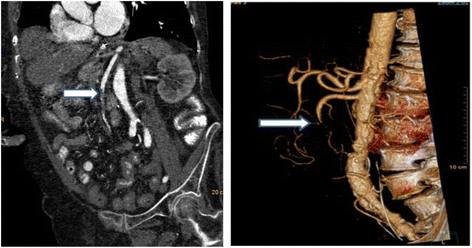
Delay in diagnosis is the dominant factor that accounts for continued mortality rates as high as 30–70% despite vast clinical experience and recognition of this entity [32, 33]. The multi-detector CTA has supplanted formal angiography as the diagnostic study of choice. Multi-detector computed tomography (MDCT) scanners are essential for the early diagnosis of AMI, but often require specialized personnel to perform and interpret the findings. 3D reconstruction is frequently helpful (Fig. 1). Volume rendering as in this image is now a semi-automatic workflow component of many CT machines. These can aide remote communities with less experienced staff.
Fig. 1
Fig. 1

Selected image from a CTA scan of a patient with acute mesenteric ischemia secondary to occluded SMA from an embolic source (arrow). 3D reconstruction is demonstrates mid occlusion of SMA (arrow)

In the presence of advanced AMI, the CTA findings reflect irreversible ischemia (intestinal dilatation and thickness, reduction or absence of visceral enhancement, pneumatosis intestinalis, and portal venous gas) and free intraperitoneal air [34].

Comprehensive biphasic CTA includes the following important steps:
  1. a)

    Pre-contrast scans to detect vascular calcification, hyper-attenuating intravascular thrombus and intramural hemorrhage.

     
  2. b)

    Arterial and venous phases to demonstrate thrombus in the mesenteric arteries and veins, abnormal enhancement of the bowel wall, and the presence of embolism or infarction of other organs.

     
  3. c)

    Multi-planar reconstructions (MPR) to assess the origin of the mesenteric arteries [35].


CTA should be performed despite the presence of renal failure, as the consequences of delayed diagnosis, missed diagnosis, or mismanagement are far more detrimental to the kidneys and the patient then exposure to the iodinated contrast agent. A recent study found that in 27 of 28 patients (96.4%) MDCT correctly diagnosed AMI (specificity of 97.9%) [16, 36]. A sensitivity of 93%, specificity of 100%, and positive and negative predictive values of 100 and 94%, respectively, were achieved [37, 38].
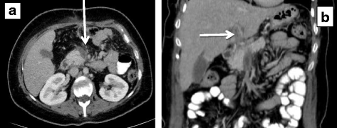
In NOMI CTA may demonstrate bowel ischemia and free fluid in the face of patent mesenteric vessels. In MVT, the most common positive radiological finding on venous phase CTA is thrombus in the superior mesenteric vein on venous phase CTA (Fig. 2). This has been described as the target sign [39].
Fig. 2
Fig. 2

30-year-old patient with acute superior mesenteric vein a and portal vein thrombosis b due to hypercoagulable state. No signs of bowel ischemia were noted, and the patient was treated successfully with long-term anticoagulation

Associated findings that suggest MVT include bowel wall thickening, pneumatosis, splenomegaly, and ascites [39]. Portal or mesenteric venous gas strongly suggests the presence of bowel infarction. Duplex ultrasonography has a limited role in this entity, but may be helpful if obtained early in chronic cases [22].

Non-occlusive mesenteric ischemia (NOMI) should be suspected in critically ill patients with abdominal pain or distension requiring vasopressor support and evidence of multi-organ dysfunction. (Recommendation 1B)

Unexplained abdominal distension or gastrointestinal bleeding may be the only signs of acute intestinal ischemia in NOMI and may be undetectable in sedated patients in the ICU in approximately 25% of cases [40, 41]. Patients surviving cardiopulmonary resuscitation who develop bacteremia and diarrhea (with or without abdominal pain) should be suspected of having NOMI. Right-sided abdominal pain associated with the passage of maroon or bright red blood in the stool is highly suggestive of NOMI in these patients.

Gastrointestinal perfusion is often impaired early in situations of critical illness, major surgery or trauma, all of which are characterized by increased demands on the circulation to maintain tissue oxygen delivery [42]. This relative mesenteric hypoperfusion is often aggravated by an underlying hypovolemic or a low-flow state. In cases of intraabdominal hypertension, all of the structures within the abdominal cavity are compressed, and this will lead to regional hypoperfusion to the organs in the splanchnic bed. Such an effect is most pronounced in the liver due to its size. Animal studies have shown that even with intraabdominal pressure of only 10 mmHg, portal venous blood flow is reduced considerably, and that at 20 mmHg, the portal venous flow and hepatic arterial flow are reduced by 35 and 55%, respectively [43].

Most of the symptoms listed in this section are often not clinically apparent in a critically ill ventilated patient on ICU. Accordingly, any negative changes in patient's physiology, including new onset of organ failure, increase in vasoactive support and nutrition intolerance should raise the suspicion of mesenteric ischemia.

When the diagnosis of AMI is made, fluid resuscitation should commence immediately to enhance visceral perfusion. Electrolyte abnormalities should be corrected, and nasogastric decompression initiated. (Recommendation 1B)

Fluid resuscitation with crystalloid and blood products is essential for the management of the patient with suspected AMI. Preoperatively resuscitation is important to prevent cardiovascular collapse on induction of anesthesia. To guide effective resuscitation, early hemodynamic monitoring should be implemented [44]. Assessment of electrolyte levels and acid–base status should be performed. This is especially true in patients with AMI, where severe metabolic acidosis and hyperkalemia may be present due to underlying bowel infarction and reperfusion [45]. Vasopressors should be used with caution, and only to avoid fluid overload and abdominal compartment syndrome. Dobutamine, low dose dopamine, and milrinone to improve cardiac function have been shown to have less impact on mesenteric blood flow [46, 47]. The fluid volume requirement in these patients may be high, due to extensive capillary leakage, but extensive crystalloid overload should be avoided to optimize bowel perfusion [48]. The endpoints of therapy should address physiologic levels of oxygen delivery with continued monitoring of lactate level as an indication of improvement. Although in the past, supra-physiologic levels were advocated, current evidence does not support this concept [49].

Broad-spectrum antibiotics should be administered immediately. Unless contraindicated, patients should be anticoagulated with intravenous unfractionacted heparin. (Recommendation 1B)

The high risk of infection among patients with AMI outweighs the risks of acquired antibiotic resistance, and therefore broad-spectrum antibiotics should be administered early in the course of treatment [50]. Intestinal ischemia leads to early loss of the mucosal barrier, which facilitates bacterial translocation and the risk of septic complications.

Prompt laparotomy should be done for patients with overt peritonitis. (Recommendation 1A)

When physical findings suggestive of an acute intraabdominal catastrophe are present, bowel infarction already occurred, and the chance of survival in this patient population with significant associated comorbidity is dramatically reduced. There is overwhelming evidence in literature that peritonitis secondary to bowel necrosis mandates surgery without delay.

The goal of surgical intervention for AMI includes:
  1. 1)

    Re-establishment blood supply to the ischemic bowel.

     
  2. 2)

    Resection of all non-viable regions.

     
  3. 3)

    Preservation of all viable bowel.


Intestinal viability is the most important factor influencing outcome in patients with AMI. Non-viable intestine, if unrecognized, results in multi-system organ dysfunction and ultimately death. Prompt laparotomy allows for direct assessment of bowel viability.

After initial resuscitation, midline laparotomy should be performed followed by assessment of all areas of the intestine with decisions for resection of all clearly necrotic areas. In cases of uncertainty, intraoperative Doppler may be helpful, as the presence of Doppler signals over distal branches of SMA facilitates bowel conservation, avoiding long-term disability. The SMA is easily palpated by placing fingers behind the root of the mesentery. The SMA is identified as a firm tubular structure, which may or may not have a palpable pulse. Otherwise, the SMA can also be reached by following the middle colic artery where it enters the SMA at the mesentery. Direct sharp dissection, exposing the artery from its surrounding mesenteric tissue, is required for proper exposure to perform revascularization. In cases of diagnostic uncertainties, arteriogram is the study of choice. It can be done intraoperatively especially in hybrid suites.
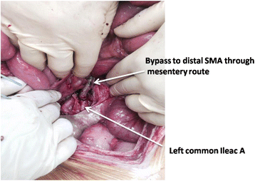
Different techniques of blood flow restoration are used depending on the pathophysiology of the AMI. Embolectomy and either primary or patch angioplasty is a well-established definitive treatment for SMA emboli. On the other hand, thrombosis of the SMA at the origin of aorta (a common pathology in diffuse aterosclerosis) will require a bypass procedure. However, it increases the magnitude of the procedure and may require prosthetics in the presence of contaminated field. One option is a retrograde bypass from the iliac artery to the distal SMA using the femoral vein or a synthetic graft (Fig. 3).
Fig. 3
Fig. 3

Patient with acute thrombosis of SMA underwent left ileo–SMA bypass with a common femoral vein graft

Neither NOMI nor MVT typically require vascular repair. Full dose anticoagulation should be initiated on all patients prior to the surgical procedure. Unfractioned heparin is effective and easy to manage, especially in patients with acute kidney failure.

Endovascular revascularization procedures may have a role with partial arterial occlusion. (Recommendation 1C)

Several case series using endovascular techniques in combination with pharmacologic therapy have been reported recently. It should be emphasized, however, that any evidence of bowel ischemia or infarction precludes the use of thrombolytic therapy. At this time, these techniques have been attempted in very early, cases of AMI, and the role of such procedures remains to be determined [51, 52]. Other contraindications to thrombolytic therapy include recent surgery, trauma, cerebrovascular or gastrointestinal bleeding, and uncontrolled hypertension [53].

In recent retrospective series of 679 patients with AMI and vascular intervention (both open and endovascular) endovascular treatment performed in 24% (165 patients). The technique was successful in 87% of the patients, and in-hospital mortality was lower than among those who underwent open procedure (25 vs. 40%) [10]. Again, this report emphasized that only patients who did not require open emergent intervention are suitable for this technical approach to revascularization.

Endovascular embolectomy may be achieved by percutaneous mechanical aspiration or thrombolysis and permits percutaneous transluminal angioplasty, with or without stenting in case series of patients with CTA evidence of acute partial or complete occlusion of the SMA (either the main trunk or branch) and without no clinical or imaging evidence of advanced bowel ischemia. Complete technical success was achieved in 28% of cases; all of these had occlusion of the main SMA trunk [54, 55, 56, 57].

There are no randomized controlled trials comparing laparotomy versus endovascular treatment as a first line strategy for the management AMI [10, 58, 59]. The most important argument in favor of the early laparotomy approach is the ability to assess bowel viability directly and thereby, minimizing delays in restoring mesenteric blood flow. In one retrospective series, the authors documented that 1/3 of patients managed with endovascular therapy avoided laparotomy [10]. In cases of endovascular approach, the use of laparoscopy to assess bowel function may be a reasonable addition [60].

Centers of excellence equipped with hybrid operating rooms may provide further data supporting the use of an endovascular strategy [61].

Damage control surgery (DCS) is an important adjunct for patients who require intestinal resection due to the necessity to reassess bowel viability and in patients with refractory sepsis. Planned re-laparotomy is an essential part of AMI management. (Recommendation 1B)

Damage control laparotomy strategy (abbreviated laparotomy) was accepted for trauma over 30 years ago and was found to be an important option in the patient with AMI. Damage control is the surgical modality of choice in the critically ill patient with AMI for physiological and technical reasons. The decision to implement the DCS mode should be made early based upon the response to resuscitation and ongoing physiology, as this has been associated with improved mortality [62]. Advanced age is not a contraindication to DCS as good outcomes have been observed in the elderly [63].

Planned second look techniques are required after restoration of SMA flow, with or without resection of ischemic bowel (and no anastomosis or stoma) following resuscitation in intensive care unit [64, 65]. Given frequent uncertainty with regard to bowel viability, the stapled off bowel ends should be left in discontinuity and re-inspected after a period of continued ICU resuscitation to restore physiological balance. Often, bowel which is borderline ischemic at the initial exploration will improve after restoration of blood supply and physiologic stabilization. Of note, however, multiple adjuncts have been suggested to assess intestinal viability, but none have proven to be uniformly reliable [66, 67].

Most often, re-exploration should be accomplished within 48 h and decisions regarding anastomosis, stoma, or additional resection can be made with plans for sequential abdominal closure.

In a review of 43 patients undergoing open mesenteric revascularization, the authors noted that 11 of the 23 patients undergoing a second-look operation required bowel resection [20]. The bowel in these patients is often very swollen and at high risk for anastomotic leak. Recent studies suggest that careful hand sewn techniques are preferable to the use of staples in this group [68, 69].

These patients often suffer from acidosis, hypothermia, and coagulation abnormalities, which require prompt and ongoing correction. Physiologic restoration is multi-factorial and includes careful and limited crystalloid infusion to avoid abdominal compartment syndrome, frequent monitoring of lactate clearance and central venous oxygen saturation as an indication of satisfactory cardiac output, and the use of viscoelastic techniques (TEG, ROTEM) to assess coagulation status and guide ongoing blood product administration. Recent evidence suggests that peritoneal resuscitation techniques may aid in this process [70, 71].

Various techniques of open abdomen have been described. The author's preferred mechanism is a simple plastic drape over the bowel, covered with a sterile towel and the use of Ioban over the abdomen. After the initial laparotomy, abdominal closure via negative pressure wound therapy is most commonly used. The open abdomen may help reduce the risk of abdominal compartment syndrome in patients requiring prolonged resuscitation. Various abdominal closure techniques have been described, however, the guiding principle is constant traction on the fascia to facilitate closure [72, 73, 74, 75].

Mesenteric venous thrombosis can often be successfully treated with a continuous infusion of unfractionated heparin. (Recommendation 1B)

MVT has a distinctive clinical finding on CTA scan, and when noted in a patient without findings of peritonitis, non-operative management should be considered. The first-line treatment for mesenteric venous thrombosis is anticoagulation. Systemic thrombolytic therapy is rarely indicated. When clinical signs demand operative intervention, one should resect only obvious necrotic bowel and employee damage control techniques liberally, since anticoagulation therapy may improve the clinical picture over the ensuing 24–48 h. Early use of heparin has been associated with improved survival [76].

Patients with peritonitis require emergency surgery. Intraoperative management is dictated by the surgical findings, which range from a segmental infarction of small bowel to necrosis of the entire bowel, with or without perforation. The aim of resection is to conserve as much bowel as possible. Second-look laparotomy, 24–48 h later, may avoid the resection of bowel that may be viable. A second-look procedure is mandatory in patients who have extensive bowel involvement.

Most published data on interventional radiological treatments for MVT are from small case series. Systemic intravenous tPA has been successfully reported [77]. Trans-jugular intrahepatic portosystemic shunt can be used for MVT with the rationale of decreasing portal pressure, which works as a vacuum of clot fragments and improves the effectiveness of thrombolysis in the case of acute thrombosis [78, 79, 80].

Supportive measures include nasogastric suction, fluid resuscitation, and bowel rest.

When NOMI is suspected, the focus is to correct the underlying cause wherever possible and to improve mesenteric perfusion. Infarcted bowel should be resected promptly. (Recommendation 1B)

Management of NOMI is based on treatment of the underlying precipitating cause. Fluid resuscitation, optimization of cardiac output, and elimination of vasopressors remain important primary measures. Additional treatment may include systemic anticoagulation and the use of catheter-directed infusion of vasodilatory and antispasmodic agents, most commonly papaverine hydrochloride [81]. The decision to intervene surgically is based on the presence of peritonitis, perforation, or overall worsening of the patient's condition [47].

If a patient presents with peritoneal signs, an exploratory laparotomy is required for resection of frankly necrotic bowel. Unfortunately, these patients are often in critical condition and the mortality remains very high (50–85%) [9]. Damage control mode is an important adjunct, given the critical state of these patients.

The finding of massive gut necrosis requires careful assessment of the patients underlying co-morbidities and advanced directives in order to judge whether comfort carries the best treatment. (Recommendation 1C)

In cases of extensive infarction of most of the small bowel with or without a portion of the colon, the surgeon could face with a philosophical decision whether to do anything. Resection of the entire involved bowel will result in short bowel syndrome with its serious associated consequences. This may not be a preferable state, particularly in elderly infirm patients, who may not tolerate long-term parenteral nutrition. A preoperative discussion with the patient and the patient's family concerning these issues is warranted and often necessary peri-operatively as well so that an agreeable plan can be reached [82].

Conclusions

AMI is a true surgical emergency. First and foremost, important evidence is a high index of suspicion based on the combination of history of abrupt onset of abdominal pain, acidosis, and organ failure. This clinical scenario should prompt imaging (CTA) in order to establish the diagnosis. In parallel with rapid resuscitation and after careful assessment of the CTA, the patient should be explored to assess bowel viability, re-establish vascular flow, and resect non-viable bowel. Subsequently, the employment of damage control techniques and continued critical care resuscitation is essential. Planned re-assessment of the bowel with further resection or anastomosis and stoma as needed is integral. Close cooperation between acute care surgeons, radiologists, anesthetists, and the vascular surgeons is essential.

Abbreviations

alpha–GST: 

Serum alpha-glutathione S-transferase

AMI: 

Acute mesenteric ischemia

 
CABA: 

Cobalt-albumin binding assay

CMI: 

Chronic mesenteric ischemia

CTA: 

Computed tomography angiography

 
DCS: 

Damage control surgery

I-FABP: 

Intestinal fatty acid binding protein

MDCT: 

Multi-detector computed tomography

MPR: 

Multi-planar reconstructions

NOMI: 

Non-occlusive mesenteric ischemia

NPWT: MVT: 

Mesenteric venous thrombosis

SMA: 

Superior mesenteric artery

 
tPA: 

Tissue plasminogen activator

WSES: 

World Society of Emergency Surgery

Declarations

Acknowledgements

Not applicable.

Funding

Not applicable.

Availability of data and materials

Not applicable.

Authors' contributions

MB, EM, and JK are responsible for the manuscript conception and draft. YK, WB, CAG, OB, CR, ZB, IC, FC, AL, AP, LA, MS, MS, SDS, GF, and FC critically revised the manuscript. All authors gave the final approval.

Ethics approval and consent to participate

Not applicable.

Consent for publication

Not applicable.

Competing interests

The authors declare that they have no competing interests.

Publisher's Note

Springer Nature remains neutral with regard to jurisdictional claims in published maps and institutional affiliations.

Open AccessThis article is distributed under the terms of the Creative Commons Attribution 4.0 International License (http://creativecommons.org/licenses/by/4.0/), which permits unrestricted use, distribution, and reproduction in any medium, provided you give appropriate credit to the original author(s) and the source, provide a link to the Creative Commons license, and indicate if changes were made. The Creative Commons Public Domain Dedication waiver (http://creativecommons.org/publicdomain/zero/1.0/) applies to the data made available in this article, unless otherwise stated.

Appendix

Current recommendations:

Statement 1

Severe abdominal pain out of proportion to physical examination findings should be assumed to be AMI until disproven. (Recommendation 1B)

Statement 2

Clinical scenario differentiates AMI as mesenteric arterial emboli, mesenteric arterial thrombosis, NOMI, or mesenteric venous thrombosis. (Recommendation 1B)

Statement 3

Conventional plain X-ray films have limited diagnostic value in evaluating AMI, although signs of intestinal perforation may be seen. (Recommendation 1B)

Statement 4

There are no laboratory studies that are sufficiently accurate to identify the presence or absence of ischemic or necrotic bowel, although elevated l-lactate and D-dimer may assist. (Recommendation 1B)

Statement 5

Computed tomography angiography (CTA) should be performed as soon as possible for any patient with suspicion for AMI. (Recommendation 1A)

Statement 6

Non-occlusive mesenteric ischemia (NOMI) should be suspected in critically ill patients with abdominal pain or distension requiring vasopressor support and evidence of multi-organ dysfunction. (Recommendation 1B)

Statement 7

When the diagnosis of AMI is made, fluid resuscitation should commence immediately to enhance visceral perfusion. Electrolyte abnormalities should be corrected, and nasogastric decompression initiated. (Recommendation 1B)

Statement 8

Broad-spectrum antibiotics should be administered immediately. Unless contraindicated, patients should be anticoagulated with intravenous unfractionated heparin. (Recommendation 1B)

Statement 9

Prompt laparotomy should be done for patients with overt peritonitis. (Recommendation 1A)

Statement 10

Endovascular revascularization procedures may have a role with partial arterial occlusion. (Recommendation 1C)

Statement 11

Damage control surgery is an important adjunct for patients who require intestinal resection due to the necessity to reassess bowel viability and in patients with refractory sepsis. Planned re-laparotomy is an essential part of AMI management. (Recommendation 1B)

Statement 12

Mesenteric venous thrombosis can often be successfully treated with a continuous infusion of unfractionated heparin. (Recommendation 1B)

Statement 13

When NOMI is suspected, the treatment focus should be to correct the underlying cause and to restore mesenteric perfusion. Infarcted bowel should be resected promptly. (Recommendation 1B)

Statement 14

The finding of massive gut necrosis requires careful assessment of the patients underlying co-morbidities and advanced directives in order to judge whether comfort carries the best treatment. (Recommendation 1C)

References

  1. Patel A, Kaleya RN, Sammartano RJ. Pathophysiology of mesenteric ischemia. Surg Clin North Am. 1992;72:31–41.View ArticlePubMedGoogle Scholar
  2. Acosta S. Mesenteric ischemia. Curr Opin Crit Care. 2015;21:171–8.View ArticlePubMedGoogle Scholar
  3. Clair DG, Beach JM. Mesenteric Ischemia. N Engl J Med. 2016;374:959–68.View ArticlePubMedGoogle Scholar
  4. Acosta S, Bjorck M. Acute thrombo-embolic occlusion of the superior mesenteric artery: a prospective study in a well-defined population. Eur J Vasc Endovasc Surg. 2003;26:179–83.View ArticlePubMedGoogle Scholar
  5. Duran M, Pohl E, Grabitz K, Schelzig H, Sagban TA, Simon F. The importance of open emergency surgery in the treatment of acute mesenteric ischemia. World J Emerg Surg. 2015;26:10–45.Google Scholar
  6. Stoney RJ, Cunningham CG. Acute mesenteric ischemia. Surgery. 1993;114:489–90.PubMedGoogle Scholar
  7. Chang RW, Chang JB, Longo WE. Update in management of mesenteric ischemia. World J Gastroenterol. 2006;12:3243–7.View ArticlePubMedPubMed CentralGoogle Scholar
  8. Horton KM, Fishman EK. Multidetector CT angiography in the diagnosis of mesenteric ischemia. Radiol Clin N Am. 2007;45:275–88.View ArticlePubMedGoogle Scholar
  9. Schoots IG, Koffeman GI, Legemate DA, Levi M, van Gulik TM. Systematic review of survival after acute mesenteric ischaemia according to disease aetiology. Br J Surg. 2004;91:17–27.View ArticlePubMedGoogle Scholar
  10. Beaulieu RJ, Arnaoutakis KD, Abularrage CJ, Efron DT, Schneider E, Black JH. Comparison of open and endovascular treatment of acute mesenteric ischemia. J Vasc Surg. 2014;59:159–64.View ArticlePubMedGoogle Scholar
  11. Rosenblum JD, Boyle CM, Schwartz LB. The mesenteric circulation. Anatomy and physiology. Surg Clin North Am. 1997;77:289–306.View ArticlePubMedGoogle Scholar
  12. Haglund U, Bergqvist D. Intestinal ischemia—the basics. Langenbeck's Arch Surg. 1999;384:233–8.View ArticleGoogle Scholar
  13. van Petersen AS, Kolkman JJ, Meerwaldt R, Huisman AB, van der Palen J, Zeebregts CJ, Geelkerken RH. Mesenteric stenosis, collaterals, and compensatory blood flow. J Vasc Surg. 2014;60:111–9.View ArticlePubMedGoogle Scholar
  14. Acosta S, Ogren M, Sternby NH, Bergqvist D, Björck M. Clinical implications for the management of acute thromboembolic occlusion of the superior mesenteric artery: autopsy findings in 213 patients. Ann Surg. 2005;241:516–22.View ArticlePubMedPubMed CentralGoogle Scholar
  15. Kärkkäinen JM, Acosta S. Acute mesenteric ischemia (part I) -incidence, etiologies, and how to improve early diagnosis. Best Pract Res Clin Gastroenterol. 2017;31:15–25.View ArticlePubMedGoogle Scholar
  16. Aschoff AJ, Stuber G, Becker BW, Hoffmann MH, Schmitz BL, Schelzig H, et al. Evaluation of acute mesenteric ischemia: accuracy of biphasic mesenteric multi-detector CT angiography. Abdom Imaging. 2009;34:345–57.View ArticlePubMedGoogle Scholar
  17. Moore HB, Moore EE, Lawson PJ, Gonzalez E, Fragoso M, Morton AP, Gamboni F, Chapman MP, Sauaia A, Banerjee A, Silliman CC. Fibrinolysis shutdown phenotype masks changes in rodent coagulation in tissue injury versus hemorrhagic shock. Surgery. 2015;158:386–92.View ArticlePubMedPubMed CentralGoogle Scholar
  18. Cohn DM, Roshani S, Middeldorp S. Thrombophilia and venous thromboembolism: implications for testing. Semin Thromb Hemost. 2007;33:573–81.View ArticlePubMedGoogle Scholar
  19. Carver TW, Vora RS, Taneja A. Mesenteric ischemia. Crit Care Clin. 2016;32:155–71.View ArticlePubMedGoogle Scholar
  20. Park WM, Gloviczki P, Cherry KJ Jr, Hallett JW Jr, Bower TC, Panneton JM, Schleck C, Ilstrup D, Harmsen WS, Noel AA. Contemporary management of acute mesenteric ischemia: Factors associated with survival. J Vasc Surg. 2002;35:445–52.View ArticlePubMedGoogle Scholar
  21. Acosta-Merida MA, Marchena-Gomez J, Hemmersbach-Miller M, Roque-Castellano C, Hernandez-Romero JM. Identification of risk factors for perioperative mortality in acute mesenteric ischemia. World J Surg. 2006;30:1579–85.View ArticlePubMedGoogle Scholar
  22. Morasch MD, Ebaugh JL, Chiou AC, Matsumura JS, Pearce WH, Yao JS. Mesenteric venous thrombosis: a changing clinical entity. J Vasc Surg. 2001;34:680–4.View ArticlePubMedGoogle Scholar
  23. Oliva IB, Davarpanah AH, Rybicki FJ, Desjardins B, Flamm SD, Francois CJ, Gerhard-Herman MD, Kalva SP, Ashraf Mansour M, Mohler ER 3rd, Schenker MP, Weiss C, Dill KE. ACR Appropriateness Criteria® imaging of mesenteric ischemia. Abdom Imaging. 2013;38:714–9.View ArticlePubMedGoogle Scholar
  24. Kougias P, Lau D, El Sayed HF, Zhou W, Huynh TT, Lin PH. Determinants ofmortality and treatment outcome following surgical interventions for acutemesenteric ischemia. J Vasc Surg. 2007;46:467–74.View ArticlePubMedGoogle Scholar
  25. Nuzzo A, Maggiori L, Ronot M, Becq A, Plessier A, Gault N, Joly F, Castier Y, Vilgrain V, Paugam C, Panis Y, Bouhnik Y, Cazals-Hatem D, Corcos O. Predictive factors of intestinal necrosis in acute mesenteric ischemia: prospective study from an intestinal stroke center. Am J Gastroenterol. 2017;112:597–605.View ArticlePubMedGoogle Scholar
  26. Evennett NJ, Petrov MS, Mittal A, Windsor JA. Systematic review and pooled estimates for the diagnostic accuracy of serological markers for intestinal ischemia. World J Surg. 2009;33:1374–83.View ArticlePubMedGoogle Scholar
  27. Powell A, Armstrong P. Plasma biomarkers for early diagnosis of acuteintestinal ischemia. Semin Vasc Surg. 2014;27:170–5.View ArticlePubMedGoogle Scholar
  28. Block T, Nilsson TK, Björck M, Acosta S. Diagnostic accuracy of plasma biomarkers for intestinal ischaemia. Scand J Clin Lab Invest. 2008;68:242–8.View ArticlePubMedGoogle Scholar
  29. Wilson C, Imrie CW. Amylase and gut infarction. Br J Surg. 1986;73:219–21.View ArticlePubMedGoogle Scholar
  30. Matsumoto S, Sekine K, Funaoka H, Yamazaki M, Shimizu M, Hayashida K, Kitano M. Diagnostic performance of plasma biomarkers in patients with acute intestinal ischaemia. Br J Surg. 2014;101:232–8.View ArticlePubMedGoogle Scholar
  31. Treskes N, Persoon AM, van Zanten ARH. Diagnostic accuracy of novel serological biomarkers to detect acute mesenteric ischemia: a systematic review and meta-analysis. Intern Emerg Med. 2017. [Epub ahead of print].Google Scholar
  32. Bradbury AW, Brittenden J, McBride K, Ruckley CV. Mesenteric ischaemia: a multidisciplinary approach. Br J Surg. 1995;82:1446–59.View ArticlePubMedGoogle Scholar
  33. Kassahun WT, Schulz T, Richter O, Hauss J. Unchanged high mortality rates from acute occlusive intestinal ischemia: six year review. Langenbeck's Arch Surg. 2008;393:163–71.View ArticleGoogle Scholar
  34. Kirkpatrick ID, Kroeker MA, Greenberg HM. Biphasic CT with mesenteric CTangiography in the evaluation of acute mesenteric ischemia: initial experience. Radiology. 2003;229:91–8.View ArticlePubMedGoogle Scholar
  35. Furukawa A, Kanasaki S, Kono N, Wakamiya M, Tanaka T, Takahashi M, Murata K. CT diagnosis of acute mesenteric ischemia from various causes. Am J Roentgenol. 2009;192:408–16.View ArticleGoogle Scholar
  36. Menke J. Diagnostic accuracy of multidetector CT in acute mesenteric ischemia: systematic review and meta-analysis. Radiology. 2010;256:93–101.View ArticlePubMedGoogle Scholar
  37. Hagspiel KD, Flors L, Hanley M, Norton PT. Computed tomography angiography and magnetic resonance angiography imaging of the mesenteric vasculature. Tech Vasc Interv Radiol. 2015;18:2–13.View ArticlePubMedGoogle Scholar
  38. Oliva IB, Davarpanah AH, Rybicki FJ, et al. ACR appropriateness criteria imaging of mesenteric ischemia. Abdom Imaging. 2013;38:714–9.View ArticlePubMedGoogle Scholar
  39. David RA, Erben Y, Kalra M. Clinical presentation, etiology, and diagnostic considerations. In: Oderich G, editor. Mesenteric Vascular Disease. New York: Springer; 2015. p. 199–209.Google Scholar
  40. Daviaud F, Grimaldi D, Dechartres A, Charpentier J, Geri G, Marin N, Chiche JD, Cariou A, Mira JP, Pène F. Timing and causes of death in septic shock. Ann Intensive Care. 2015;5:16.View ArticlePubMedPubMed CentralGoogle Scholar
  41. Guillaume A, Pili-Floury S, Chocron S, Delabrousse E, De Parseval B, Koch S, Samain E, Capellier G, Piton G. Acute mesenteric ischemia among post-cardiac surgery patients presenting with multiple organ failure. Shock. 2016. [Epub ahead of print].Google Scholar
  42. Mastoraki A, Mastoraki S, Tziava E, Touloumi S, Krinos N, Danias N, Lazaris A, Arkadopoulos N. Mesenteric ischemia: pathogenesis and challenging diagnostic and therapeutic modalities. World J Gastrointest Pathophysiol. 2016;7:125–30.View ArticlePubMedPubMed CentralGoogle Scholar
  43. Mogilner JG, Bitterman H, Hayari L, et al. Effect of elevated intra-abdominal pressure and hyperoxia on portal vein blood flow, hepatocyte proliferation and apoptosis in a rat model. Eur J Pediatr Surg. 2008;18:380–6.View ArticlePubMedGoogle Scholar
  44. Wyers MC. Acute mesenteric ischemia: diagnostic approach and surgical treatment. Semin Vasc Surg. 2010;23:9–20.View ArticlePubMedGoogle Scholar
  45. Corcos O, Nuzzo A. Gastro-intestinalvascular emergencies. Best Pract Res Clin Gastroenterol. 2013;27:709–25.View ArticlePubMedGoogle Scholar
  46. Tendler DA, LaMont JT. Acute mesenteric ischemia. Uptodate. 2013. http://www.uptodate.com/contents/acute-mesentericischemia. Accessed 13 Aug 2016.
  47. Sise MJ. Mesenteric ischemia: the whole spectrum. Scand J Surg. 2010;99:106–10.View ArticlePubMedGoogle Scholar
  48. Oldenburg WA, Lau LL, Rodenberg TJ, Edmonds HJ, Burger CD. Acute mesentericischemia: a clinical review. Arch Intern Med. 2004;164:1054–62.View ArticlePubMedGoogle Scholar
  49. Helmerhorst HJ, Schultz MJ, van der Voort PH, de Jonge E, van Westerloo DJ. Bench-to-bedside review: the effects of hyperoxia during critical illness. Crit Care. 2015;19:284.View ArticlePubMedPubMed CentralGoogle Scholar
  50. Silvestri L, van Saene HK, Zandstra DF, Marshall JC, Gregori D, Gullo A. Impactof selective decontamination of thedigestive tract on multiple organ dysfunctionsyndrome: systematic review of randomizedcontrolled trials. Crit Care Med. 2010;38:1370–6.View ArticlePubMedGoogle Scholar
  51. Schoots IG, Levi MM, Reekers JA, Lameris JS, van Gulik TM. Thrombolytic therapy for acute superior mesenteric artery occlusion. J Vasc Interv Radiol. 2005;16:317–29.View ArticlePubMedGoogle Scholar
  52. Landis MS, Rajan DK, Simons ME, Hayeems EB, Kachura JR, Sniderman KW. Percutaneous management of chronic mesenteric ischemia: outcomes after intervention. J Vasc Interv Radiol. 2005;16:1319–25.View ArticlePubMedGoogle Scholar
  53. Sarac TP. Techniques of endovascular revascularization for acute mesenteric ischemia. In: Oderich G, editor. Mesenteric Vascular Disease. New York: Springer; 2015. p. 241–52.Google Scholar
  54. Kim BG, Ohm JY, Bae MN, Kim HN, Kim YJ, Chung MH, Park CS, Ihm SH, Kim HY. Successful percutaneous aspiration thrombectomy for acute mesenteric ischemia in a patient with atrial fibrillation despite optimal anticoagulation therapy. Can J Cardiol. 2013;29:1329.e5–7.View ArticleGoogle Scholar
  55. Schoenbaum SW, Pena C, Koenigsberg P, Katzen BT. Superior mesenteric artery embolism: treatment with intra arterial urokinase. JVIR. 1992;3:485–90.View ArticlePubMedGoogle Scholar
  56. Simó G, Echenagusia AJ, Camuñez F, Turégano F, Cabrera A, Urbano J. Superior mesenteric arterial embolism: local fibrinolytic treatment with urokinase. Radiology. 1997;204:775–9.View ArticlePubMedGoogle Scholar
  57. Jia Z, Jiang G, Tian F, Zhao J, Li S, Wang K, Wang Y, Jiang L, Wang W. Early endovascular treatment of superior mesenteric occlusion secondary to thromboemboli. Eur J Vasc Endovasc Surg. 2014;47:196–203.View ArticlePubMedGoogle Scholar
  58. Orr NT, Endean ED. Part two: against the motion. an endovascular first strategy is not the optimal approach for treating acute mesenteric ischemia. Eur J Vasc Endovasc Surg. 2015;50:276–9.View ArticlePubMedGoogle Scholar
  59. Arthurs ZM, Titus J, Bannazadeh M, Eagleton MJ, Srivastava S, Sarac TP, et al. A comparison of endovascular revascularization with traditional therapy for the treatment of acute mesenteric ischemia. J Vasc Surg. 2011;53:698–705.View ArticlePubMedGoogle Scholar
  60. Cocorullo G, Mirabella A, Falco N, Fontana T, Tutino R, Licari L, Salamone G, Scerrino G, Gulotta G. An investigation of bedside laparoscopy in the ICU for cases of non-occlusive mesenteric ischemia. World J Emerg Surg. 2017;12:4.View ArticlePubMedPubMed CentralGoogle Scholar
  61. Björck M. Part one: for the motion. an endovascular first strategy is the optimal approach for treating acute mesenteric ischemia. Eur J Vasc Endovasc Surg. 2015;50:273–5.View ArticlePubMedGoogle Scholar
  62. Godat L, Kobayashi L, Costantini T, Coimbra R. Abdominal damage control surgery and reconstruction: world society of emergency surgery position paper. World J Emerg Surg. 2013;8:53.View ArticlePubMedPubMed CentralGoogle Scholar
  63. Weber DG, Bendinelli C, Balogh ZJ. Damage control surgery for abdominal emergencies. Br J Surg. 2014;101:109–18.View ArticleGoogle Scholar
  64. Subramanian A, Balentine C, Palacio CH, Sansgiry S, Berger DH, Awad SS. Outcomes of damage-control celiotomy in elderly non trauma patients with intra-abdominal catastrophes. Am J Surg. 2010;200:783–8.View ArticlePubMedGoogle Scholar
  65. Weber DG, Bendinelli C, Balogh ZJ. Damage control surgery for abdominal emergencies. Br J Surg. 2014;101:e109–18.View ArticlePubMedGoogle Scholar
  66. Urbanavičius L, Pattyn P, de Putte DV, Venskutonis D. How to assess intestinal viability during surgery: a review of techniques. World J Gastrointest Surg. 2011;3:59–69.View ArticlePubMedPubMed CentralGoogle Scholar
  67. Horgan PG, Gorey TF. Operative assessment of intestinal viability. Surg Clin North Am. 1992;72:143–55.View ArticlePubMedGoogle Scholar
  68. Worrell S, Mumtaz S, Tsuboi K, Lee TH, Mittal SK. Anastomotic complications associated with stapled versus hand-sewn anastomosis. J Surg Res. 2010;161:9–12.View ArticlePubMedGoogle Scholar
  69. Brundage SI, Jurkovich GJ, Hoyt DB, Patel NY, Ross SE, Marburger R, Stoner M, Ivatury RR, Ku J, Rutherford EJ, Maier RV, WTA Multi-institutional Study Group, Western Trauma Association. Stapled versus sutured gastrointestinal anastomoses in the trauma patient: a multicenter trial. J Trauma. 2001;51:1054–61.View ArticlePubMedGoogle Scholar
  70. Smith JW, Matheson PJ, Franklin GA, Harbrecht BG, Richardson JD, Garrison RN. Randomized controlled trial evaluating the efficacy of peritoneal resuscitation in the management of trauma patients undergoing damage control surgery. J Am Coll Surg. 2017;224:396–404.View ArticlePubMedGoogle Scholar
  71. Crafts TD, Hunsberger EB, Jensen AR, Rescorla FJ, Yoder MC, Markel TA. Direct peritoneal resuscitation improves survival and decreases inflammation after intestinal ischemia and reperfusion injury. J Surg Res. 2015;199:428–34.View ArticlePubMedGoogle Scholar
  72. Acosta S, Björck M, Petersson U. Vacuum-assisted wound closure and mesh-mediated fascial traction for open abdomen therapy—a systematic review. Anaesthesiol Intensive Ther. 2017.[Epub ahead of print].Google Scholar
  73. Hofmann AT, Gruber-Blum S, Lechner M, Petter-Puchner A, Glaser K, Fortelny R. Delayed closure of open abdomen in septic patients treated with negative pressure wound therapy and dynamic fascial suture: the long-term follow-up study. Surg Endosc. 2017. [Epub ahead of print].Google Scholar
  74. Willms A, Schaaf S, Schwab R, Richardsen I, Jänig C, Bieler D, Wagner B, Güsgen C. Intensive care and health outcomes of open abdominal treatment: long-term results of vacuum-assisted wound closure and mesh-mediated fascial traction (VAWCM). Langenbeck's Arch Surg. 2017;402:481–92.View ArticleGoogle Scholar
  75. Tolonen M, Mentula P, Sallinen V, Rasilainen S, Bäcklund M, Leppäniemi A. Open abdomen with vacuum-assisted wound closure and mesh-mediated fascial traction in tients with complicated diffuse secondary peritonitis: A single-center 8-year experience. J Trauma Acute Care Surg. 2017;82:1100–5.View ArticlePubMedGoogle Scholar
  76. Acosta S. Surgical management of peritonitis secondary to acute superior mesenteric artery occlusion. World J Gastroenterol. 2014;20:9936–41.View ArticlePubMedPubMed CentralGoogle Scholar
  77. Hmoud B, Singal AK, Kamath PS. Mesenteric venous thrombosis. J Clin Exp Hepatol. 2014;4:257–63.View ArticlePubMedPubMed CentralGoogle Scholar
  78. al Karawi MA, Quaiz M, Clark D, Hilali A, Mohamed AE, Jawdat M. Mesenteric vein thrombosis, non-invasive diagnosis and follow-up (US + MRI), and non-invasive therapy by streptokinase and anticoagulants. Hepato-Gastroenterology. 1990;37:507–9.PubMedGoogle Scholar
  79. Semiz-Oysu A, Keussen I, Cwikiel W. Interventional radiological management of prehepatic obstruction of [corrected] the splanchnic venous system. Cardiovasc Intervent Radiol. 2007;30:688–95.View ArticlePubMedGoogle Scholar
  80. Sehgal M, Haskal ZJ. Use of transjugular intrahepatic portosystemic shunts during lytic therapy of extensive portal splenic and mesenteric venous thrombosis: long-term follow-up. J Vasc Interv Radiol. 2000;11:61–5.View ArticlePubMedGoogle Scholar
  81. Meilahn JE, Morris JB, Ceppa EP, Bulkley GB. Effect of prolonged selective intramesenteric arterial vasodilator therapy on intestinal viability after acute segmental mesenteric vascular occlusion. Ann Surg. 2001;234:107–15.View ArticlePubMedPubMed CentralGoogle Scholar
  82. Lilley EJ, Cooper Z, Schwarze ML, Mosenthal AC. Palliative care in surgery: defining the research priorities. J Palliat Med. 2017;20:702–9.View ArticlePubMedGoogle Scholar

Copyright

© The Author(s). 2017
Alexandros Sfakianakis
Anapafseos 5 . Agios Nikolaos
Crete.Greece.72100
2841026182
6948891480

Annales françaises d'Oto-rhino-laryngologie et de Pathologie Cervico-faciale

Une tumeur sous-muqueuse de la base de langue

Publication date: April 2019

Source: Annales françaises d'Oto-rhino-laryngologie et de Pathologie Cervico-faciale, Volume 136, Issue 2

Author(s): A. Ko, C.-L. Hyun, G.-C. Lim


Vésicules endobuccales, pharyngées et laryngées unilatérales

Publication date: April 2019

Source: Annales françaises d'Oto-rhino-laryngologie et de Pathologie Cervico-faciale, Volume 136, Issue 2

Author(s): L. Alciato, P. Bonfils, F. Rubin


Les pharyngotomies latérales

Publication date: April 2019

Source: Annales françaises d'Oto-rhino-laryngologie et de Pathologie Cervico-faciale, Volume 136, Issue 2

Author(s): O. Laccourreye, A. Villeneuve, F. Rubin, C.F. Holsinger

Résumé

Dans cette note technique, les auteurs précisent, au vu des données de la littérature médicale scientifique, les points techniques clefs, les variantes, les erreurs à éviter, ainsi que les principaux résultats fonctionnels des pharyngotomies latérales dans les cancers dont le point de départ se situe au niveau de l'oro et/ou de l'hypopharynx.


Chirurgie de l'hypophyse par abord transnasal transseptal sous guidage endoscopique

Publication date: April 2019

Source: Annales françaises d'Oto-rhino-laryngologie et de Pathologie Cervico-faciale, Volume 136, Issue 2

Author(s): V. Favier, J. Boetto, C. Cartier, F. Segnarbieux, L. Crampette

Résumé

La chirurgie de l'hypophyse est réalisée par voie transsphénoïdale dans l'immense majorité des cas. Les méthodes ont varié au fil du temps. Les voies transseptales par une incision muqueuse sous-labiale ou par une incision muqueuse nasale, sous contrôle microscopique, ont été largement utilisées. L'usage de l'endoscope par voie transnasale a été décrit dans les années 1990. Il s'est ensuivi la conception que l'abord transseptal était attaché à l'usage du microscope et que l'abord endoscopique se faisait par la voie endonasale stricte. Nous utilisons la voie transseptale transsphénoïdale, par abord muqueux nasal, intégralement guidée par endoscopie, tant pour l'abord que pour l'exérèse. Cette technique présente à nos yeux de nombreux avantages : abord strictement médian de la selle turcique, large champ opératoire, absence de conflit d'instruments et peu complications nasales.


Lipomes cervicaux symptomatiques

Publication date: April 2019

Source: Annales françaises d'Oto-rhino-laryngologie et de Pathologie Cervico-faciale, Volume 136, Issue 2

Author(s): Y. Najaf, C. Cartier, V. Favier, R. Garrel

Résumé
Introduction

Les lipomes sont des tumeurs bénignes fréquentes, que l'on retrouve dans l'ensemble du corps humain ; une minorité d'entre elles est située au niveau de la tête et du cou.

Résumé du cas clinique

Dans cet article, nous décrivons trois différents cas illustrés de lipomes cervicaux symptomatiques. Les diagnostics différentiels et les thérapeutiques y sont discutés à la lumière de la littérature médicale sur ce sujet.

Discussion

Les lipomes cervicaux ont en règle générale une croissance lente et peuvent être responsables de symptômes divers tels qu'une dysphagie, une dyspnée ou une dysphonie liées à un effet de masse, ou bien être uniquement révélés par une gêne esthétique. Il est important de garder en mémoire le potentiel de transformation maligne des lipomes, qui représente un challenge diagnostique. Dans cet article, nous avons essayé de mettre en évidence la nécessité d'un dialogue entre chirurgiens, anatomopathologistes et radiologues pour le diagnostic et la prise en charge des lipomes de la tête et du cou.


Récidive de lichen plan hypertrophique du méat auditif externe

Publication date: April 2019

Source: Annales françaises d'Oto-rhino-laryngologie et de Pathologie Cervico-faciale, Volume 136, Issue 2

Author(s): A. Košec, M. Kostić, J. Ajduk, M. Ries

Résumé
Introduction

Nous rapportons un cas de lichen plan hypertrophique primitif évolutif unilatéral du méat auditif externe ayant nécessité plusieurs interventions chirurgicales pour traiter un prurit permanent, une otorrhée, une sténose et une hypoacousie de transmission.

Résumé du cas

Patiente de 58 ans initialement traitée par méatoplastie pour suspicion d'otite externe oblitérante chronique. Après un intervalle libre asymptomatique de 5 ans, la maladie a récidivé localement avec atteinte d'autres surfaces corporelles. L'otorrhée, l'hypoacousie de transmission et le prurit se sont aggravés, et une mastoïdectomie en technique ouverte a été réalisée, avec résection complète de la peau du méat auditif externe et de la membrane tympanique. Le diagnostic de lichen plan a été confirmé par l'analyse histopathologique.

Discussion

Très peu de résultats chirurgicaux concernant la sténose du méat auditif externe causée par le lichen plan ont été publiés. Un décollement et une exérèse complète de la peau de la portion médiale du méat auditif externe, suivie d'une greffe dermo-épidermique sont recommandés en traitement initial. Nous discutons des options thérapeutiques et du résultat chirurgical après échec de la chirurgie initiale.


Sinusite invasive à Scedosporium Apiospermum révélée par un abcès extradural

Publication date: April 2019

Source: Annales françaises d'Oto-rhino-laryngologie et de Pathologie Cervico-faciale, Volume 136, Issue 2

Author(s): N. Khoueir, B. Verillaud, P. Herman

Résumé
Introduction

La rhinosinusite fongique invasive chronique (RSFIC) est une entité rare qui affecte généralement les sujets immunodéprimés. L'agent pathogène le plus fréquemment retrouvé est Aspergillus. Elle prend le plus souvent à l'imagerie la forme d'une pathologie agressive pseudotumorale. Nous présentons un cas de RSFIC à Scedosporium Apiospermum (S. Apiospermum) avec une présentation clinique et radiologique atypique.

Résumé du cas

Un homme immunocompétent âgé de 72 ans présentait des céphalées chroniques, des cervicalgies et une limitation bilatérale dans le regard latéral. L'imagerie révélait une pathologie sphénoïdale gauche isolée avec des remaniements osseux importants et un abcès extradural en regard du clivus. Une sphénoïdotomie endoscopique large avec rhinopharyngectomie de type II était réalisée et le diagnostic de RSFIC à S. Apiospermum était posé sur les données de l'histologie et de la culture fongique. Le patient refusait tout traitement médical. Il n'y avait pas de récidive après 1 an de suivi.

Discussion

S. Apiospermum est un germe rarement isolé dans la RSIFC. Notre cas était révélé par une présentation clinique atypique incluant une infection sphénoïdale isolée compliquée par une paralysie bilatérale du VI et un abcès extradural. L'imagerie était également inhabituelle, prenant plutôt l'aspect d'une dysplasie fibreuse ou d'une ostéomyélite bactérienne que l'aspect pseudotumoral typique. Le patient était traité avec succès par chirurgie seule, qui pourrait donc être suffisante chez les sujets immunocompétents.


Épiglottopexie par puncture transcutanée pour prolapsus de l'épiglotte en cas de laryngomalacie sévère. Une nouvelle technique

Publication date: April 2019

Source: Annales françaises d'Oto-rhino-laryngologie et de Pathologie Cervico-faciale, Volume 136, Issue 2

Author(s): H. Álvarez-Neri, P. Villamor, E. Ortiz, J.-P. Grub

Résumé
Introduction

La laryngomalacie (LM) est la première cause de stridor chez le nourrisson et le jeune enfant. Chez 10 à 20 % des patients atteints de LM, une chirurgie peut être nécessaire en cas de symptômes sévères. La supraglottoplastie est la chirurgie la plus répandue pour traiter la LM sévère mais elle ne suffit pas en cas de bascule rostrocaudale de l'épiglotte contre la paroi pharyngée postérieure.

Résumé du cas clinique

Nous rapportons le cas d'un nourrisson de 2 mois atteint de laryngomalacie sévère avec un collapsus glottique majeur de l'épiglotte entraînant une obstruction secondaire des voies aériennes, des troubles de la déglutition et un retard de croissance staturopondérale. Une épiglottopexie par puncture transcutanée a été réalisée avec un résultat satisfaisant. Avec un recul de 2 ans, le patient est resté asymptomatique sans aucun événement indésirable.

Discussion

L'obstruction glottique secondaire à un collapsus postérieur de l'épiglotte est le type de laryngomalacie le plus sévère entraînant des symptômes respiratoires graves et un retard de croissance staturopondérale. L'épiglottopexie par puncture transcutanée est une nouvelle technique, peu onéreuse car elle ne nécessite pas de matériel spécifique et peut être réalisée par laryngoscopie en suspension. Elle peut être pratiquée seule ou en association avec une supraglottoplastie traditionnelle.


Lipomatose cervico-dorsale révélant un syndrome MERRF (myoclonic epilepsy with ragged red fibers)

Publication date: April 2019

Source: Annales françaises d'Oto-rhino-laryngologie et de Pathologie Cervico-faciale, Volume 136, Issue 2

Author(s): F. Carré, R. Hervochon, C. Foirest, F. Tankéré

Résumé
Introduction

Les patients atteints d'un syndrome MERRF (myoclonic epilepsy with ragged red fibers) présentent principalement une encéphalo-myopathie. La lipomatose, rarement présente, est de localisation cervico-dorsale, progressive et récidivante.

Cas clinique

Nous rapportons 4 cas cliniques présentant l'association d'une lipomatose au syndrome MERRF. Chez 3 patients, le diagnostic de syndrome MERRF a pu être réalisé du fait de la présentation clinique lipomateuse et grâce à l'interrogatoire retrouvant des antécédents personnels ou familiaux de lipomes et de myopathie.

Discussion

La présence d'une lipomatose axiale extensive doit faire rechercher d'autres signes cliniques du syndrome MERRF chez le patient ou sa famille. Les enjeux diagnostiques sont liés au conseil génétique.


Recommandations de la SFORL (version courte) sur les malformations lymphatiques cervicales de l'adulte et de l'enfant : le diagnostic

Publication date: April 2019

Source: Annales françaises d'Oto-rhino-laryngologie et de Pathologie Cervico-faciale, Volume 136, Issue 2

Author(s): J. Lerat, A. Bisdorff-Bresson, M. Borsic, C. Chopinet, V. Couloignier, N. Fakhry, P. Fayoux, F. Jegoux, A. Larralde, N. Leboulanger, R. Nicollas, S. Pondaven Letourmy, Groupe de travail de la SFORL

Résumé
Objectif

Les auteurs exposent les recommandations de la SFORL concernant le diagnostic des malformations lymphatiques cervicales de l'adulte et de l'enfant.

Méthodes

À partir de la revue de la littérature scientifique et de l'expérience d'un groupe de travail multidisciplinaire, des recommandations ont été rédigées, puis relues par un groupe de lecture indépendant, et finalisées dans une réunion de coordination. Les recommandations proposées ont été classées en grade A, B, C ou accord professionnel selon un niveau de preuve scientifique décroissant.

Résultats

Il est recommandé de faire un examen clinique ORL complet afin d'identifier les lésions à risque de complications ou de mauvais pronostic. Il est recommandé, en cas de doute diagnostique pour les lésions latéro-cervicales et du plancher buccal, de réaliser une cytoponction avant une décision thérapeutique. Il est recommandé d'utiliser une ou plusieurs des classifications validées pour évaluer l'efficacité des traitements et suivre l'évolution de la maladie. Il est recommandé, afin d'améliorer la fiabilité du diagnostic anténatal d'associer IRM et échographie. Il est recommandé dans le cadre du diagnostic anténatal d'évaluer de façon précise l'extension locorégionale d'une malformation lymphatique cervicale afin d'en estimer le pronostic, et de rechercher systématiquement des malformations associées afin de guider les propositions thérapeutiques.


Alexandros Sfakianakis
Anapafseos 5 . Agios Nikolaos
Crete.Greece.72100
2841026182
6948891480

Oral Biology

BMP4 mutations in tooth agenesis and low bone mass

Publication date: July 2019

Source: Archives of Oral Biology, Volume 103

Author(s): Miao Yu, Hao Wang, Zhuangzhuang Fan, Chencheng Xie, Haochen Liu, Yang Liu, Dong Han, Sing-Wai Wong, Hailan Feng

Abstract
Objective

To identify an uncommon genetic cause of tooth agenesis (TA) by utilizing whole exome sequencing (WES) and targeted Sanger sequencing in a cohort of 120 patients with isolated TA.

Design

One deleterious mutation in the gene encoding bone morphogenetic protein 4 (BMP4) was identified in 6 unrelated patients with TA by WES. After that, the coding exons of BMP4 were examined in 114 TA patients using Sanger sequencing. Dual-energy X-ray absorptiometry (DEXA) was used to measure the bone mineral density of patients who carried a BMP4 mutation. Finally, preliminary functional studies of two BMP4 mutants were performed.

Results

We detected 3 novel missense mutations (c.58 G > A: p.Gly20Ser, c.326 G > T: p.Arg109Leu and c.614 T > C: p.Val205Ala) and 1 reported mutation in the BMP4 gene among 120 TA probands. The previously reported BMP4 mutation (c.751C > T: p.His251Tyr) was associated with urethra and eye anomalies. By extending the pedigrees, we determined that the tooth phenotypes had an autosomal dominant inheritance pattern, as individuals carrying a BMP4 mutation exhibit different types of dental anomalies. Interestingly, we observed that patients harboring a BMP4 mutation manifested early onset osteopenia or osteoporosis. Further in vitrofunctional assays demonstrated that two BMP4 mutants resulted in a decreased activation of Smad signaling. Therefore, a loss-of-function in BMP4 may contribute to the clinical phenotypes seen in this study.

Conclusions

We identified 4 mutations in the BMP4 gene in 120 TA patients. To our knowledge, this is the first study to describe human skeletal diseases associated with BMP4 mutations.


Interactions among moderate/severe periodontitis, ADIPOQ-rs1501299, and LEPR-rs1137100 polymorphisms on the risk of type 2 diabetes in a Chinese population

Publication date: July 2019

Source: Archives of Oral Biology, Volume 103

Author(s): Xiaojing Cao, Pengcheng Huo, Wenjing Li, Peng Li, Lu He, Huanxin Meng

Abstract
Objective

Type 2 diabetes mellitus (T2DM) is a complex disease influenced by genes and the environment. Periodontitis a demonstrated risk factor of T2DM. Previous studies related to gene-environment interactions on the risk of T2DM mainly focused on gene-obesity interactions. However, the impact of gene-periodontitis interaction on the risk of T2DM has not yet been investigated. This study aimed to investigate gene-environment interactions among moderate/severe periodontitis, polymorphisms of adiponectin (ADIPOQ)-rs1501299, and leptin receptor (LEPR)-rs1137100 on T2DM risk in Chinese subjects.

Design

A case-control study was conducted in 239 Chinese participants from Beijing Hypertension Association Institute (BHAL). After full-mouth periodontal examinations, the participants underwent bilateral buccal swabs for DNA testing. ADIPOQ-rs1501299 and LEPR-rs1137100 were used for genotyping. Generalised multifactor dimensionality reduction (GMDR) and logistic regression were used to examine the interactions among single nucleotide polymorphisms (SNPs) and moderate/severe periodontitis on the risk of T2DM.

Results

The risk of T2DM was higher in moderate/severe periodontitis [adjusted odds ratio (AOR) = 3.67, 95% confidence interval (95%CI): 1.26–10.71] in ADIPOQ-rs1501299 GG genotype (AOR = 3.42, 95%CI: 1.81–6.46) and LEPR-rs1137100 GG genotype (AOR = 3.16, 95%CI: 1.56–6.39). The GMDR model indicated that there was a significant three-factor model (p = 0.001) involving rs1501299, rs1137100, and moderate/severe periodontitis, demonstrating a potential gene-environment interaction among periodontitis, polymorphisms of rs1501299, and rs1137100 influencing the risk of T2DM. Moderate/severe periodontitis patients with rs1501299-GG and rs1137100-GG have the highest T2DM risk after adjusting for age, gender, BMI, WHR, smoking status, alcohol consumption, economic status, and hypertension (AOR = 20.39, 95%CI: 2.64–157.26).

Conclusions

Interactions among moderate/severe periodontitis, rs1501299-GG, and rs1137100-GG were associated with an increased risk of T2DM. This study may provide a new insight into the effect of gene-environment interactions on T2DM.


Effect of analogues of cationic peptides on dentin mineralization markers in odontoblast-like cells

Publication date: July 2019

Source: Archives of Oral Biology, Volume 103

Author(s): Karina S. Caiaffa, Fernanda G. Basso, Norival A. Santos-Filho, Carlos Alberto de Souza-Costa, Vivien T. Sakai, Eduardo M. Cilli, Cristiane Duque

Abstract
Objectives

To evaluate the effect of analogues of cationic peptides on the viability and the expression of phenotypic and genotypic markers of dentin mineralization in MDPC-23 odontoblast-like cells.

Materials and methods

Cells were exposed to serial dilutions of analogues of cationic peptides hBD-3-1CV and KR-12-a5 compared to peptide LL-37 and their viability was assessed by methyltetrazolium assay. Next, peptides (0.78–62.5 μg/mL) were applied on the MDPC-23 cells for evaluating the total protein (TP) production, alkaline phosphatase (ALP) activity and mineralized nodule deposition. Gene expression of mineralization markers (DSPP and DMP-1) was also determined by quantitative PCR.

Results

LL-37 and hBD-3-1CV treatment did not affect cellular viability at concentrations below 62.5 μg/mL. KR-12-a5 reduced cell viability above 31.25 μg/mL. TP production was similar for all groups compared with the control group, except by hBD-3-1CV (at 15.62 μg/mL). LL-37 (at 62.5 μg/mL) induced higher ALP activity than control and other experimental groups. LL-37 and hBD-3-1CV, at 62.5 μg/mL and KR-12-a5 at 31.25 μg/mL stimulated the highest deposition of mineralized nodule. Overall, no statistical differences were observed between the groups for DSPP-1 and DMP-1 expressions.

Conclusions

LL-37 was the only peptide that induced both ALP activity and mineralized nodules deposition, without affecting cell viability. None of peptides tested induced the expression of DSPP or DMP-1, genes commonly involved in active dentin mineralization.


Tooth agenesis-related GLI2 and GLI3 genes may contribute to craniofacial skeletal morphology in humans

Publication date: July 2019

Source: Archives of Oral Biology, Volume 103

Author(s): Guido Artemio Marañón-Vásquez, Beatriz Dantas, Christian Kirschneck, Juliana Arid, Arthur Cunha, Alice Gomes de Carvalho Ramos, Marjorie Ayumi Omori, Amanda Silva Rodrigues, Ellen Cardoso Teixeira, Simone Carvalho Levy, Agnes Schroeder, Mírian Aiko Nakane Matsumoto, Peter Proff, Lívia Azeredo A. Antunes, Alexandre R. Vieira, Leonardo Santos Antunes, Erika Calvano Küchler

Abstract
Objective

The present cross-sectional, multi-centre, genetic study aimed to determine, whether single nucleotide polymorphisms (SNPs) in tooth agenesis (TA)-associated GLI2 and GLI3 genes contribute to the development of craniofacial skeletal morphology in humans.

Design

Orthodontic patients from an ethnically heterogeneous population were selected for the present study (n = 594). The presence or absence of TA was determined by analysis of panoramic radiography and dental records. The subjects were classified according to their skeletal malocclusion and facial growth pattern by means of digital cephalometric analysis. Genomic DNA was extracted from squamous epithelial cells of the buccal mucosa and SNPs in GLI2 (rs3738880, rs2278741) and GLI3 (rs929387, rs846266) were analysed by polymerase chain reaction using TaqMan chemistry and end-point analysis.

Results

Class II skeletal malocclusion presented a significantly lower frequency of TA (P < 0.05). Subjects without TA showed significantly higher ANB angles (P < 0.05). Genotype and/or allele distributions of the SNPs in GLI2(rs3738880, rs2278741) and GLI3 (rs846266) were associated with the presence of TA (P < 0.05). The SNPs rs3738880, rs2278741 and rs929387 were also associated with some type of skeletal malocclusion (P < 0.05), but not with the facial growth pattern (P > 0.05). The G allele for TA-related GLI2 rs3738880 was strongly linked to the presence of Class III skeletal malocclusion (OR = 2.03; 95% CI = 1.37–3.03; P<3125 × 10−6). GLI2rs2278741 C allele was overrepresented in individuals without TA, suggesting it as a protective factor for this dental phenotype (OR = 0.43; 95% CI = 0.24−0.78; P<625 × 10−5).

Conclusion

The present study suggests that SNPs in TA-associated GLI2 and GLI3 genes may also play a role in the development of skeletal malocclusions. rs3738880 and rs2278741 in GLI2 seems to contribute to the genetic background for skeletal Class III and TA, respectively. TA could be an additional predictor of craniofacial morphology in some cases. Further research replicating the reported associations should be performed.


WNT10A mutations causing oligodontia

Publication date: July 2019

Source: Archives of Oral Biology, Volume 103

Author(s): Haemin Park, Ji-Soo Song, Teo Jeon Shin, Hong-Keun Hyun, Young-Jae Kim, Jung-Wook Kim

Abstract
Objectives

To identify the molecular genetic etiology of the families with non-syndromic multiple missing permanent teeth (oligodontia).

Materials and methods

Genomic DNA was isolated and measured, and whole-exome sequencing was performed. The obtained sequencing reads were aligned to the human reference genome and subsequently processed by a series of bioinformatics programs. Finally, short insertions/deletions and single nucleotide variations were annotated with dbSNP build 138.

Results

The proband of family 1 was missing 14 permanent teeth, and the mutational analysis revealed compound heterozygousWNT10A mutations (c.364A > T and c.511C > T). Two affected individuals in family 2 were missing 20 and 12 permanent teeth, respectively, and compound heterozygous WNT10A mutations (c.364A > T and c.637G > A) were also identified.

Conclusions

This study reveals compound heterozygousWNT10A missense mutations in two families with non-syndromic oligodontia which will improve the understanding of odontogenesis and the pathogenesis related to WNT10Amutations.


The effect of vanillic acid on ligature-induced periodontal disease in Wistar rats

Publication date: July 2019

Source: Archives of Oral Biology, Volume 103

Author(s): Ozkan Karatas, Hatice Balci Yuce, Mehmet Murat Taskan, Fikret Gevrek, Fatma Ucan Yarkac, Aslı Keskin, Sukruye Firuze Ocak Karatas, Hulya Toker

Abstract
Objective

Vanillic acid, also known as 4-hydroxy-3-methoxy benzoic acid has a potent effect on bone metabolism. The purpose of the present study was to specify the effects of vanillic acid (VA) on preventing inflammation and bone destruction in experimental periodontitis as inflammatory bone disease. To evaluate the effects of VA, osteoblast, osteoclast and inflammatory cell counts, iNOS, CD68, MMP-1, and TIMP-1 levels were determined.

Methods

32 female Wistar rats were divided into four experimental groups as; Group 1: healthy control (C, n = 8), group 2: Periodontitis (P, n = 8), group 3: periodontitis and 50 mg/kg VA administered group (P + VA-50, n = 8) and group 4: periodontitis and 100 mg/kg VA delivered group (P + VA-100, n = 8). Ligature-induced experimental periodontitis was carried out at mandibular first molar teeth of the right quadrant by placing submarginal 4-0 silk ligatures. VA was administered by oral gavage for 14 days beginning from the first day. Rats were euthanized on the 15th day. Morphological changes in alveolar bone were evaluated via a stereomicroscope. Mandibles were subjected to histological procedures. Osteoblasts, tartrate-resistant acid phosphatase synthesizing osteoclasts and inflammatory cells were counted. Inducible nitric oxide synthase (iNOS), cluster of differentiation (CD)-68, Matrix metalloproteinase (MMP)-1, tissue inhibitor of MMP-1, runt-related x factor-2 (RUNX2), and cyclooxygenase (COX)-2 expressions were determined by immunohistochemistry.

Results

The rats in the periodontitis group had the highest alveolar bone loss compared to the other groups. Both doses of VA significantly decreased alveolar bone loss but not the control levels. TRAP-positive osteoclast and inflammatory cell counts were also highest in the P group, and both 50 and 100 mg/kg VA reduced these counts. Control rats had the lowest osteoclast and inflammatory cell counts compared to the other groups. Similar to osteoclast counts, MMP-1, iNOS, CD68, and COX-2 expressions were the highest in the P group compared to the other groups. Both doses of VA significantly decreased these levels. Osteoblast cells were higher in the VA groups compared to the control and periodontitis groups. RUNX2 levels were lower in the periodontitis group compared to the control group. A slight increase was also observed in VA groups. However, the difference in the TIMP-1 levels was significant only between P and VA100 groups.

Conclusion

VA administration successfully ameliorated periodontitis symptoms by decreasing alveolar bone and collagen destruction, periodontal inflammation, and increasing osteoblastic activity.


Interleukin-17 plays a role in pulp inflammation partly by WNT5A protein induction

Publication date: July 2019

Source: Archives of Oral Biology, Volume 103

Author(s): Mengyu Liu, Yuan Zhao, Chenglin Wang, Haiyun Luo, Peng A, Ling Ye

Abstract
Objective

Our study aimed to investigate the role of interleukin (IL)-17 in dental pulp inflammation and the relationship between WNT5A and IL-17.

Methods

Immunohistochemical staining was used to detect the expression of tumor necrosis factor-α (TNF-α), WNT5A and IL-17 in pulp tissues. Anti-IL-17 neutralizing antibody was used in rat pulpitis model and to study the role of IL-17 in pulpitis. TNF-α, WNT5A or IL-17 recombinant protein were used to treat human dental pulp cells. RT-PCR, Western blot, and Enzyme linked immunosorbent assay were used to detect the expression of mRNA and protein. Transwell assay was used to measure the migration of THP-1 cells, which is a human monocytic cell line.

Results

IL-17 and WNT5A are co-expressed in TNF-α high-expressed region in human and rat pulpitis tissue. IL-17 mainly contributes to its positive regulatory role in inflammation through up regulate cytokines and mediated macrophages migration. Anti-IL-17 neutralizing antibody can suppress the inflammatory cell infiltration and TNF-α expression in dental pulpitis. TNF-α promotes the expression of IL-17 partly through WNT5A and WNT5A regulates IL-17 expression by mitogen-activated protein kinase (MAPK)-(P38 and ERK) pathway.

Conclusions

IL-17 acts as an inflammatory mediator in dental pulp inflammation. The expression of IL-17 can be partially regulated by WNT5A.


Effects of 7S globulin 3 derived from the adzuki bean [Vigna angularis] on the CSP- and eDNA- dependent biofilm formation of Streptococcus mutans

Publication date: June 2019

Source: Archives of Oral Biology, Volume 102

Author(s): Hidenobu Senpuku, Shota Mohri, Mamiko Mihara, Toshiaki Arai, Yusuke Suzuki, Yoji Saeki

Abstract
Objective

Streptococcus mutans is a principal bacterium that forms pathogenic biofilm involved in the development of dental caries. S. mutans possesses a quorum sensing system (QS) stimulated by competence stimulating peptide (CSP), which is associated with bacteriocin production, genetic competency and biofilm formation. Inhibiting CSP-dependent QS is one of the aims leading to the inhibition of biofilm formation and is useful for establishing new prevention systems for dental caries.

Design

In this study, we selected adzuki bean [Vigna angularis] extract as a candidate component to inhibit CSP-dependent biofilm formation among various foods. To purify an inhibitory component from the adzuki extracts, we performed the salting-out method, two rounds of ion-exchange chromatography, and SDS and native PAGE.

Results

A primary protein band that inhibits CSP-dependent biofilm formation appeared at approximately 50 kDa and was identified as 7S globulin 3 (7S3), a major seed storage protein in adzuki bean. To determine the characteristics of 7S3 as an inhibitory component, aggregated proteins were extracted from the adzuki crude extracts at pH values lower than 6. The aggregated proteins inhibited CSP- and eDNA-dependent biofilm formation and showed 50 kDa band, which is identical with 7S3 in the purified sample. Moreover, 7S globulin 3 in the adzuki bean extract directly interacted with CSP at low pH conditions but not at neutral conditions, and inhibited CSP-dependent bacteriocin production.

Conclusion

It was suggested that 7S3 might be a safe and useful material to prevent pathogenic activities in the biofilm formation of S. mutans.


Occlusal interference induces oxidative stress and increases the expression of UCP3 in the masseter muscle: A rat model

Publication date: June 2019

Source: Archives of Oral Biology, Volume 102

Author(s): Donglei Wu, Jing Liu

Abstract
Objective

To determine whether occlusal alteration contributes to masticatory muscle damage by inducing oxidative stress.

Design

Thirty Sprague-Dawley rats were randomly divided into six groups, including occlusal interference groups (3 days, 7 days, 14 days, 21 days, and removal for 3 days) and a sham group. A rat experimental model of occlusal interference was generated by a 0.6-mm unilateral bite-raise. The rats were euthanised for evaluation of histologic changes in the masseter muscles using haematoxylin-eosin staining. To further investigate the role of oxidative stress and uncoupling protein (UCP3) in the development of occlusal dysfunction-induced masseter damage, levels of UCP3 protein were measured by western blot analysis.

Results

Compared with the sham group, the connective tissue of the masseter muscle was extended partially and inflammatory cells appeared following the induction of malocclusion. With respect to the oxidative stress markers, there were increases in malondialdehyde (MDA) content but decreases in superoxide dismutase (SOD) and glutathione peroxidase (GPX) activities; furthermore, the expression of UCP3 was upregulated. After eliminating the occlusal interference for 3 days, the degree of inflammation was substantially alleviated, the MDA content decreased, and SOD and GPX activities increased. The expression of UCP3 decreased.

Conclusions

Occlusal interference induces oxidative stress in the masseter muscle, regulated by UCP3. Overall, these findings have significant implications for the understanding of how occlusal dysfunction causes muscle fatigue and pain.


Effect of dioxin-related compounds on oral pigmentation in patients affected by the Yusho incident

Publication date: June 2019

Source: Archives of Oral Biology, Volume 102

Author(s): Goro Kawasaki, Izumi Yoshitomi

Abstract
Background/aim

Toxins such as polychlorinated biphenyls (PCBs) and dioxins dramatically affect patients even decades after exposure. Patients with Yusho, a condition caused by exposure to PCBs and dioxins, have diverse mental and physical complaints, even though it is almost 50 years since the Yusho incident. Oral pigmentation is one of the major symptoms in Yusho patients.

Patients and methods

A total of 183 participants in the Yusho health study were examined. Oral examinations, including recording the prevalence of oral pigmentation, were performed by two oral surgeons. Demographic and clinical characteristics, including blood concentration of PCB and 2,3,4,7,8-pentachlorodibezofuran (2,3,4,7,8-PeCDF), which are the major causes of Yusho, were obtained from the results of recent surveys conducted by the Yusho Study Group.

Results

The mean serum PCB and 2,3,4,7,8-PeCDF levels of the 183 Yusho patients were 1.59 ± 1.25 ppb and 29.0 ± 42.9 pg/g lipid, respectively. There was a significant correlation between the levels of PCB and 2,3,4,7,8-PeCDF (r = 0.64, p < 0.01). The rate of oral pigmentation in Yusho patients (25.7%) was significant higher than among potential victims of Yusho (13 of 183, 7.1%) (p < 0.05).

Conclusion

The prevalence of oral pigmentation was still significantly higher in Yusho patients, even 50 years after exposure, although blood PCB levels have decreased in that time.


Alexandros Sfakianakis
Anapafseos 5 . Agios Nikolaos
Crete.Greece.72100
2841026182
6948891480

NeuroImage

Shared neural representations of syntax during online dyadic communication

Publication date: September 2019

Source: NeuroImage, Volume 198

Author(s): Wenda Liu, Holly P. Branigan, Lifen Zheng, Yuhang Long, Xialu Bai, Kanyu Li, Hui Zhao, Siyuan Zhou, Martin J. Pickering, Chunming Lu

Abstract

When people communicate, they come to see the world in a similar way to each other by aligning their mental representations at such levels as syntax. Syntax is an essential feature of human language that distinguishes humans from other non-human animals. However, whether and how communicators share neural representations of syntax is not well understood. Here we addressed this issue by measuring the brain activity of both communicators in a series of dyadic communication contexts, by using functional near-infrared spectroscopy (fNIRS)-based hyperscanning. Two communicators alternatively spoke sentences either with the same or with different syntactic structures. Results showed a significantly higher-level increase of interpersonal neural synchronization (INS) at right posterior superior temporal cortex when communicators produced the same syntactic structures as each other compared to when they produced different syntactic structures. These increases of INS correlated significantly with communication quality. Our findings provide initial evidence for shared neural representations of syntax between communicators.


Multimodal characterization of the human nucleus accumbens

Publication date: September 2019

Source: NeuroImage, Volume 198

Author(s): Samuel CD. Cartmell, Qiyuan Tian, Brandon J. Thio, Christoph Leuze, Li Ye, Nolan R. Williams, Grant Yang, Gabriel Ben-Dor, Karl Deisseroth, Warren M. Grill, Jennifer A. McNab, Casey H. Halpern

Abstract

Dysregulation of the nucleus accumbens (NAc) is implicated in numerous neuropsychiatric disorders. Treatments targeting this area directly (e.g. deep brain stimulation) demonstrate variable efficacy, perhaps owing to non-specific targeting of a functionally heterogeneous nucleus. Here we provide support for this notion, first observing disparate behavioral effects in response to direct simulation of different locations within the NAc in a human patient. These observations motivate a segmentation of the NAc into subregions, which we produce from a diffusion-tractography based analysis of 245 young, unrelated healthy subjects. We further explore the mechanism of these stimulation-induced behavioral responses by identifying the most probable subset of axons activated using a patient-specific computational model. We validate our diffusion-based segmentation using evidence from several modalities, including MRI-based measures of function and microstructure, human post-mortem immunohistochemical staining, and cross-species comparison of cortical-NAc projections that are known to be conserved. Finally, we visualize the passage of individual axon bundles through one NAc subregion in a post-mortem human sample using CLARITY 3D histology corroborated by 7T tractography. Collectively, these findings extensively characterize human NAc subregions and provide insight into their structural and functional distinctions with implications for stereotactic treatments targeting this region.


The relation between brain signal complexity and task difficulty on an executive function task

Publication date: September 2019

Source: NeuroImage, Volume 198

Author(s): John G. Grundy, Ryan M. Barker, John A.E. Anderson, Judith M. Shedden

Abstract

On a daily basis, we constantly deal with changing environmental cues and perceptual conflicts and as such, our brains must flexibly adapt to current demands in order to act appropriately. Brains become more efficient and are able to switch states more readily by increasing the complexity of their neural networks. However, it is unclear how brain signal complexity relates to behavior in young adults performing cognitively demanding executive function tasks. Here we used multiscale entropy analysis and multivariate statistics on EEG data while participants performed a bivalency effect task-switching paradigm to show that brain signal complexity in young adults increases as task demands increase, that increases in brain signal complexity are associated with both speed gains and losses depending on scalp location, and that more difficult tasks are associated with more circumscribed complexity across the scalp. Overall, these findings highlight a critical role for brain signal complexity in predicting behavior on an executive function task among young adults.


Group-level cortical and muscular connectivity during perturbations to walking and standing balance

Publication date: September 2019

Source: NeuroImage, Volume 198

Author(s): Steven M. Peterson, Daniel P. Ferris

Abstract

Maintaining balance is a complex process requiring multisensory processing and coordinated muscle activation. Previous studies have indicated that the cortex is directly involved in balance control, but less information is known about cortical flow of signals for balance. We studied source-localized electrocortical effective connectivity dynamics of healthy young subjects (29 subjects: 14 male and 15 female) walking and standing with both visual and physical perturbations to their balance. The goal of this study was to quantify differences in group-level corticomuscular connectivity responses to sensorimotor perturbations during walking and standing. We hypothesized that perturbed visual input during balance would transiently decrease connectivity between occipital and parietal areas due to disruptive visual input during sensory processing. We also hypothesized that physical pull perturbations would increase cortical connections to central sensorimotor areas because of higher sensorimotor integration demands. Our findings show decreased occipito-parietal connectivity during visual rotations and widespread increases in connectivity during pull perturbations focused on central areas, as expected. We also found evidence for communication from cortex to muscles during perturbed balance. These results show that sensorimotor perturbations to balance alter cortical networks and can be quantified using effective connectivity estimation.


Change detection to tone pairs during the first year of life – Predictive longitudinal relationships for EEG-based source and time-frequency measures

Publication date: September 2019

Source: NeuroImage, Volume 198

Author(s): Jarmo A. Hämäläinen, Silvia Ortiz-Mantilla, April Benasich

Abstract

Brain responses related to auditory processing show large changes throughout infancy and childhood with some evidence that the two hemispheres might mature at different rates. Differing rates of hemispheric maturation could be linked to the proposed functional specialization of the hemispheres in which the left auditory cortex engages in analysis of precise timing information whereas the right auditory cortex focuses on analysis of sound frequency. Here the auditory change detection process for rapidly presented tone-pairs was examined in a longitudinal sample of infants at the age of 6 and 12 months using EEG. The ERP response related to change detection of a frequency contrast, its estimated source strength in the auditory areas, as well as time-frequency indices showed developmental effects. ERP amplitudes, source strength, spectral power and inter-trial phase locking decreased across age. A differential lateralization pattern emerged between 6 and 12 months as shown by inter-trial phase locking at 2–3 Hz; specifically, a larger developmental change was observed in the right as compared to the left hemisphere. Predictive relationships for the change in source strength from 6 months to 12 months were found. Six-month predictors were source strength and phase locking values at low frequencies. The results show that the infant change detection response in rapidly presented tone pairs is mainly determined by low frequency power and phase-locking with a larger phase-locking response at 6 months predicting greater change at 12 months. The ability of the auditory system to respond systematically across stimuli is suggested as a marker of maturational change that leads to more automatic and fine-tuned cortical responses.


Enhancing neural efficiency of cognitive processing speed via training and neurostimulation: An fNIRS and TMS study

Publication date: September 2019

Source: NeuroImage, Volume 198

Author(s): Adrian Curtin, Hasan Ayaz, Yingying Tang, Junfeng Sun, Jijun Wang, Shanbao Tong

Abstract

Speed of Processing (SoP) represents a fundamental limiting step in cognitive performance which may underlie General Intelligence. The measure of SoP is particularly sensitive to aging, neurological or cognitive diseases, and has become a benchmark for diagnosis, cognitive remediation, and enhancement. Neural efficiency of the Dorsolateral Prefrontal Cortex (DLPFC) is proposed to account for individual differences in SoP. However, the mechanisms by which DLPFC efficiency is shaped by training and whether it can be enhanced remain elusive. To address this, we monitored the brain activity of sixteen healthy participants using functional Near Infrared Spectroscopy (fNIRS) while practicing a common SoP task (Symbol Digit Substitution Task) across 4 sessions. Furthermore, in each session, participants received counterbalanced excitatory repetitive transcranial magnetic stimulation (rTMS) during mid-session breaks. Results indicate a significant involvement of the left-DLPFC in SoP, whose neural efficiency is consistently increased through task practice. Active neurostimulation, but not Sham, significantly enhanced the neural efficiency. These findings suggest a common mechanism by which neurostimulation may aid to accelerate learning.


Topological correction of infant white matter surfaces using anatomically constrained convolutional neural network

Publication date: September 2019

Source: NeuroImage, Volume 198

Author(s): Liang Sun, Daoqiang Zhang, Chunfeng Lian, Li Wang, Zhengwang Wu, Wei Shao, Weili Lin, Dinggang Shen, Gang Li, UNC/UMN Baby Connectome Project Consortium

Abstract

Reconstruction of accurate cortical surfaces without topological errors (i.e., handles and holes) from infant brain MR images is very important in early brain development studies. However, infant brain MR images typically suffer extremely low tissue contrast and dynamic imaging appearance patterns. Thus, it is inevitable to have large amounts of topological errors in the segmented infant brain tissue images, which lead to inaccurately reconstructed cortical surfaces with topological errors. To address this issue, inspired by recent advances in deep learning, we propose an anatomically constrained network for topological correction on infant cortical surfaces. Specifically, in our method, we first locate regions of potential topological defects by leveraging a topology-preserving level set method. Then, we propose an anatomically constrained network to correct those candidate voxels in the located regions. Since infant cortical surfaces often contain large and complex handles or holes, it is difficult to completely correct all errors using one-shot correction. Therefore, we further enroll these two steps into an iterative framework to gradually correct large topological errors. To the best of our knowledge, this is the first work to introduce deep learning approach for topological correction of infant cortical surfaces. We compare our method with the state-of-the-art methods on both simulated topological errors and real topological errors in human infant brain MR images. Moreover, we also validate our method on the infant brain MR images of macaques. All experimental results show the superior performance of the proposed method.

Graphical abstract

Image 1


Empathy to emotional voices and the use of real-time fMRI to enhance activation of the anterior insula

Publication date: September 2019

Source: NeuroImage, Volume 198

Author(s): Dana Kanel, Salim Al-Wasity, Kristian Stefanov, Frank E. Pollick

Abstract

The right anterior insula (AI), known to have a key role in the processing and understanding of social emotions, is activated during tasks that involve the act of empathising. Neurofeedback provides individuals with a visualisation of their own brain activity, enabling them to regulate and modify this activity. Following previous research investigating the ability of individuals to up-regulate right AI activity levels through neurofeedback, we investigated whether this could be similarly accomplished during an empathy task involving auditory stimuli of human positive and negative emotional expressions. Twenty participants, ten with feedback from right anterior insula and ten with feedback from a sham brain region, participated in two sessions that included sixteen neurofeedback runs and four transfer runs. Results showed that for the second session participants in the right AI neurofeedback group demonstrated better ability to up-regulate their right AI compared to the control group who received sham feedback. Examination of the relationship between individual participants' empathic traits and their ability to up-regulate right AI activity showed that participants low on empathic traits produced a greater increase in activation of right AI by the end of training. Moreover, the response to positively valenced audio stimuli was greater than for negatively valenced stimuli. These results have implications for therapeutic training of empathy in populations with limited empathic response.


Microelectrode array electrical impedance tomography for fast functional imaging in the thalamus

Publication date: September 2019

Source: NeuroImage, Volume 198

Author(s): Danyi Zhu, Alistair McEwan, Calvin Eiber

Abstract

Electrical Impedance Tomography (EIT) has the potential to be able to observe functional tomographic images of neural activity in the brain at millisecond time-scales. Prior modelling and experimental work has shown that EIT is capable of imaging impedance changes from neural depolarisation in rat somatosensory cortex. Here, we investigate the feasibility of EIT for imaging impedance changes using a stereotaxically implanted microelectrode array in the thalamus. Microelectrode array EIT was simulated using an anatomically accurate marmoset brain model. Impedance imaging was validated and detectability estimated using physiological noise recorded from the marmoset visual thalamus. The results suggest that visual-input-driven impedance changes in visual subcortical bodies within 300 μm of the implanted array could be reliably reconstructed and localised, comparable to local field potential measurements. Furthermore, we demonstrated that microelectrode array EIT could reconstruct concurrent activity in multiple subcortical bodies simultaneously.

Graphical abstract

Image 1


Rhythmicity facilitates pitch discrimination: Differential roles of low and high frequency neural oscillations

Publication date: September 2019

Source: NeuroImage, Volume 198

Author(s): Andrew Chang, Dan J. Bosnyak, Laurel J. Trainor

Abstract

Previous studies indicate that temporal predictability can enhance timing and intensity perception, but it is not known whether it also enhances pitch perception, despite pitch being a fundamental perceptual attribute of sound. Here we investigate this in the context of rhythmic regularity, a form of predictable temporal structure common in sound streams, including music and speech. It is known that neural oscillations in low (delta: 1–3 Hz) and high (beta: 15–25 Hz) frequency bands entrain to rhythms in phase and power, respectively, but it is not clear why both low and high frequency bands entrain to external rhythms, and whether they and their coupling serve different perceptual functions. Participants discriminated near-threshold pitch deviations (targets) embedded in either rhythmic (regular/isochronous) or arrhythmic (irregular/non-isochronous) tone sequences. Psychophysically, we found superior pitch discrimination performance for target tones in rhythmic compared to arrhythmic sequences. Electroencephalography recordings from auditory cortex showed that delta phase, beta power modulation, and delta-beta coupling were all modulated by rhythmic regularity. Importantly, trial-by-trial neural-behavioural correlational analyses showed that, prior to a target, the depth of U-shaped beta power modulation predicted pitch discrimination sensitivity whereas cross-frequency coupling strength predicted reaction time. These novel findings suggest that delta phase might reflect rhythmic temporal expectation, beta power temporal attention, and delta-beta coupling auditory-motor communication. Together, low and high frequency auditory neural oscillations reflect different perceptual functions that work in concert for tracking rhythmic regularity and proactively facilitate pitch perception.


Alexandros Sfakianakis
Anapafseos 5 . Agios Nikolaos
Crete.Greece.72100
2841026182
6948891480

Cardiothoracic Anesthesia

Pressure-regulated volume control mode versus synchronized intermittent mandatory ventilation mode in management of acute respiratory failure complicating advanced liver disease
Rafik Yousset Atalla, Dalia Fahmy Emam, Wail Ahmed Abdelaal

The Egyptian Journal of Cardiothoracic Anesthesia 2018 12(3):35-41

Background A common cause of death in the ICUs is advanced liver disease and its complications. Respiratory complications are common in patients with advanced liver disease. Management of patients with advanced hepatic disease having acute respiratory failure mandates the use of lung-protective ventilation strategies to achieve adequate oxygenation. Objectives The aim of this study was to compare and evaluate the superiority of either pressure-regulated volume control (PRVC) mode versus synchronized intermittent mandatory ventilation (SIMV) mode in management of acute respiratory failure complicating advanced hepatic disease. Patients and methods A total of 80 patients were included in this study, who were randomized into two equal groups of 40 patients each, namely. group S and group PV. Group S was ventilated using SIMV mode, whereas group PV was ventilated using PRVC mode. Results There was a statistically significant difference between group PV and group S during the mechanical ventilation period regarding the PO2/FiO2 ratio, static compliance, and dynamic compliance values, with higher values in group PV than group S providing better oxygenation. There was a statistically significant difference in the duration of mechanical ventilation and the ICU stay between the two groups, with significantly lower values in the PV group than the S group. Conclusion We concluded that PRVC mode of mechanical ventilation is superior to SIMV in acute respiratory failure complicating advanced liver disease, as it resulted in improved oxygenation at lower inflation pressures. 


Intraoperative thromboelastographic assessment of platelet function in a patient with low platelet count and high-mean platelet volume requiring cardiac surgery
Francesco Del Sindaco, Daniel Rajeev, Sandra Faloye, Lisa Vipond

The Egyptian Journal of Cardiothoracic Anesthesia 2018 12(3):42-44

An 80-year-old patient with low platelet count was admitted in our institution for aortic valve replacement and a coronary artery bypass graft. Because of thrombocytopenia, a mean platelet volume has been assessed, showing a value above the range. Two pools of platelets were booked but not transfused, neither in the theater nor during the postoperative period. A thromboelastographic assessment at the end of the cardiopulmonary bypass showed a normal overall platelet function, and no clinical issues (diffuse bleeding after protamine and/or oozing from the drain above the range, in the postoperative period) were noticed. The authors point out the importance of thromboelastographic or thromboelastometric assessments in patients with isolated thrombocytopenia and high-mean platelet volume to contribute to reduce the risk of inappropriate transfusions. 


Tamponade-like picture caused by pericardial cyst
Mostafa Eladawy, Anup Varghese

The Egyptian Journal of Cardiothoracic Anesthesia 2018 12(3):45-48

Pericardial cysts are rare entities, they have an incidence rate of 1:100,000 which constitutes about 7.6% of all mediastinal masses (6). The differential diagnosis includes other solid tumors and cysts of the mediastinum, diaphragmatic hernia or tumors, aneurysms of the heart or great vessels (1). Although TTE can differentiate many of the causes of mediastinal masses, loculated pericardial effusion with a similar clinical picture can be difficult to differentiate on transthoracic echocardiography (8). Radiological examination with CT or MRI has been described to differentiate in difficult cases. In emergency situations, TEE examination could be a useful option as it can help differentiating a malignant from a benign pericardial cyst, check the extent of involvement of underlying myocardium or adjacent structures and assess the anatomical abnormalities or effects on different heart chambers. In our case we describe a case of a huge pericardial cyst which had affected the patient haemodynamics in a tamponade like picture, the intra-operative TEE assessment helped appreciating the compression effect on the right heart chambers and the intravascular resuscitation needed to counteract the external pressure of the pericardial cyst. The echocardiography also helped the decision making of performing the procedure on CPB. 


Alexandros Sfakianakis
Anapafseos 5 . Agios Nikolaos
Crete.Greece.72100
2841026182
6948891480

Αρχειοθήκη ιστολογίου