Acta Radiologica RSS feed -- OnlineFirst Articles
Feasibility study of reduced field of view diffusion-weighted magnetic resonance imaging in head and neck tumors
Vidiri, A., Minosse, S., Piludu, F., Curione, D., Pichi, B., Spriano, G., Marzi, S. · Saturday, June 11, 2016, 7:57
Background
Reduced field of view (rFOV) imaging may be used to improve the quality of diffusion-weighted imaging (DWI) in the head and neck (HN) region.
Purpose To evaluate the feasibility of rFOV-DWI in patients affected by HN tumors, through a comparison with conventional full FOV (fFOV) DWI.
Material and Methods Twenty-two patients with histologically-proven malignant or benign tumors of the head and neck were included in a retrospective study. All patients underwent pre-treatment magnetic resonance imaging (MRI) studies including rFOV-DWI and fFOV-DWI. The apparent diffusion coefficient (ADC) value distributions inside tumor and muscle were derived and the mean, standard deviation (SD), and kurtosis were calculated. Image distortion was quantitatively and qualitatively evaluated, as well as the capability of lesion identification. The Wilcoxon test was used to compare all variables. Agreements between the ADC estimations were assessed by Bland–Altman plots.
Results Image distortion and lesion identification scores were both higher for rFOV-DWI compared to fFOV-DWI. A reduction in ADC values with rFOV-DWI emerged for both lesion and muscle, with a mean percentage difference in ADC of 6.2% in the lesions and 24.9% in the muscle. The difference in SD of ADC was statistically significant in the lesions, indicating a higher ADC homogeneity for rFOV DWI (P = 0.005).
Conclusion The application of rFOV DWI in patients affected by HN tumors is feasible and promising, based on both qualitative and quantitative analyses. This technique has potential for improving the diagnostic accuracy of fFOV-DWI for the study of specific tumoral areas.
Tags: Imaging
Acta Radiologica RSS feed -- OnlineFirst Articles
Patellar position in weight-bearing radiographs compared with non-weight-bearing: significance for the detection of osteoarthritis
Skou, N., Egund, N. · Saturday, June 11, 2016, 7:57
Background
Diagnosis and treatment of patellofemoral disorders including osteoarthritis are currently often based on imaging and clinical assessment with patients in the supine position.
Purpose To evaluate differences in patellar position in the trochlear groove and to assess the detection of medial and lateral patellofemoral (PF) osteoarthritis (OA) on axial radiographs in supine and standing positions, respectively.
Material and Methods Thirty-five women and 23 men (mean age, 56 years; age range, 18–87 years) referred for routine radiographic examinations of the knees were included. Axial radiographs of the PF joint in both supine non-weight-bearing and standing weight-bearing position in 30° knee flexion were obtained of 111 knees. Measurements performed on the radiographs: patellar tilt, patellar displacement, joint space width, and grade of OA according to Ahlbäck.
Results From supine to standing position the patella moved medially and medial joint space width and lateral patellar tilt angle decreased (P < 0.0001 for the three measured parameters). In the standing position, medial PF OA was observed in 19 knees compared to three knees in the supine position. Fourteen knees had lateral PF OA with almost unchanged grade of OA irrespective of position.
Conclusion In weight-bearing positions, the patella is positioned medially in the trochlear groove compared to supine non-weight-bearing positions. Therefore, this study suggests that the common occurrence of medial PF OA can generally not be detected on axial radiographs in supine non-weight-bearing positions and confirms the importance of imaging the PF joint in standing weight-bearing positions.
Tags: Imaging
Clinical Imaging
Usefulness of breath-hold inversion recovery-prepared T1-weighted two-dimensional gradient echo sequence for detection of hepatocellular carcinoma in Gd-EOB-DTPA-enhanced MR imaging
Friday, June 10, 2016, 20:34
Publication date: September–October 2016
Source:Clinical Imaging, Volume 40, Issue 5
Author(s): Tsuyoshi Ohno, Hiroyoshi Isoda, Akihiro Furuta, Shigeki Arizono, Rikiya Yamashita, Ayako ono, Kaori Togashi
The aim is to evaluate the diagnostic performance and the added value of breath-hold inversion recovery-prepared T1-weighted two-dimensional gradient echo (IR-2D-GRE) sequence for detection of hepatocellular carcinoma (HCC) in patients with insufficient liver parenchymal enhancement during the hepatobiliary phase (HBP) of Gd-EOB-DTPA-enhanced magnetic resonance imaging (MRI). Seventeen patients with a quantitative liver-to-spleen contrast ratio of ≤1.5 on HBP images and 36 HCCs were included. Liver-to-lesion contrast ratios on HBP images obtained with IR-2D-GRE sequence were significantly higher than those with three-dimensional gradient echo sequence. The addition of IR-2D-GRE sequence during HBP of Gd-EOB-DTPA-enhanced MRI yielded higher diagnostic accuracy and improved sensitivity.
Tags: Imaging
Neuroradiology
Functional connectivity change of brain default mode network in breast cancer patients after chemotherapy
Friday, June 10, 2016, 7:22
Abstract
Introduction
Complaint about attention disorders is common among breast cancer patients who have undergone chemotherapy, which may be associated with the default mode network (DMN). To validate this hypothesis, we investigated the DMN functional connectivity (FC) change and its relationship with the attention function in breast cancer patients (BC) using resting-state functional magnetic resonance imaging (rs-fMRI).
Methods
Twenty-two BC treated with chemotherapy and 22 healthy controls (HC) were recruited into this study. The FC between the DMN's hubs and regions of the dorsal medial prefrontal cortex (dMPFC) and medial temporal lobe (MTL) subsystems was respectively calculated for each participant.
Results
The statistical result showed significantly lower connectivity in dMPFC and MTL subsystems in the BC group. In addition, the partial correlation analysis result indicated that the low connectivity of some brain regions in MTL subsystem was correlated with attention dysfunction following BC chemotherapy.
Conclusion
These results suggest that the functional disconnection in MTL subsystem of the DMN may have association with attention function of BC after chemotherapy.
Tags: Imaging, Neurology
Medical Image Analysis
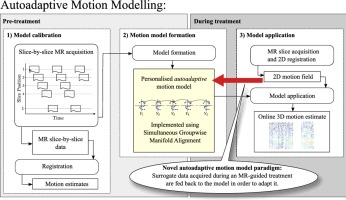
Autoadaptive Motion Modelling for MR-Based Respiratory Motion Estimation
Friday, June 10, 2016, 7:04
Publication date: Available online 9 June 2016
Source:Medical Image Analysis
Author(s): Christian F. Baumgartner, Christoph Kolbitsch, Jamie R. McClelland, Daniel Rueckert, Andrew P. King
Respiratory motion poses significant challenges in image-guided interventions. In emerging treatments such as MR-guided HIFU or MR-guided radiotherapy, it may cause significant misalignments between interventional road maps obtained pre-procedure and the anatomy during the treatment, and may affect intra-procedural imaging such as MR-thermometry. Patient specific respiratory motion models provide a solution to this problem. They establish a correspondence between the patient motion and simpler surrogate data which can be acquired easily during the treatment. Patient motion can then be estimated during the treatment by acquiring only the simpler surrogate data. In the majority of classical motion modelling approaches once the correspondence between the surrogate data and the patient motion is established it cannot be changed unless the model is recalibrated. However, breathing patterns are known to significantly change in the time frame of MR-guided interventions. Thus, the classical motion modelling approach may yield inaccurate motion estimations when the relation between the motion and the surrogate data changes over the duration of the treatment and frequent recalibration may not be feasible. We propose a novel methodology for motion modelling which has the ability to automatically adapt to new breathing patterns. This is achieved by choosing the surrogate data in such a way that it can be used to estimate the current motion in 3D as well as to update the motion model. In particular, in this work, we use 2D MR slices from different slice positions to build as well as to apply the motion model. We implemented such an autoadaptive motion model by extending our previous work on manifold alignment. We demonstrate a proof-of-principle of the proposed technique on cardiac gated data of the thorax and evaluate its adaptive behaviour on realistic synthetic data containing two breathing types generated from 6 volunteers, and real data from 4 volunteers. On synthetic data the autoadaptive motion model yielded 21.45% more accurate motion estimations compared to a non-adaptive motion model 10 minutes after a change in breathing pattern. On real data we demonstrated the method's ability to maintain motion estimation accuracy despite a drift in the respiratory baseline. Due to the cardiac gating of the imaging data, the method is currently limited to one update per heart beat and the calibration requires approximately 12 minutes of scanning. Furthermore, the method has a prediction latency of 800 ms. These limitations may be overcome in future work by altering the acquisition protocol.
Graphical abstract
Tags: BioEngineering, Imaging
Pediatric Radiology
Searching for certainty: findings predictive of appendicitis in equivocal ultrasound exams
Friday, June 10, 2016, 3:58
Abstract
Background
Ultrasound (US) is the preferred imaging modality for evaluating suspected pediatric appendicitis. However, borderline appendiceal enlargement or questionable inflammatory changes can confound interpretation and lead to equivocal exams.
Objective
The purpose of this study was to determine which findings on equivocal US exams are most predictive of appendicitis.
Materials and methods
All US exams performed for suspected pediatric appendicitis from July 1, 2013, through July 9, 2014, were initially interpreted using a risk-stratified scoring system. Two blinded pediatric radiologists independently reviewed US exams designated as equivocal and recorded the following findings: increased wall thickness, loss of mural stratification, peri-appendiceal fat inflammation, peri-appendiceal fluid, appendicolith and maximum appendiceal diameter. A third pediatric radiologist resolved discrepancies. US features were correlated with the final diagnosis via multivariate analysis.
Results
During the study period, 162/3,750 (4.3%) children had US exams initially interpreted as equivocal (mean age 9.8 +/- 3.8 years). Five outpatients were lost to follow-up. Forty-eight of the remaining 157 (30.6%) children had an operative diagnosis of appendicitis. Findings significantly associated with appendicitis were loss of mural stratification (odds ratio [OR] = 6.7, P=0.035), peri-appendiceal fat inflammation (OR = 10.0, P<0.0001) and appendicolith (OR = 15.8, P=0.025). While appendiceal diameter tended to be larger in patients with appendicitis, the difference was not statistically significant.
Conclusion
Loss of mural stratification, peri-appendiceal fat inflammation and an appendicolith are significant predictors of appendicitis in children with otherwise equivocal US exams. While maximum appendiceal diameter is not statistically associated with appendicitis in our study, mean appendiceal diameter of 6.7 mm in those without appendicitis suggests that the customary upper normal limit of 6 mm is too sensitive.
Tags: Imaging
Pediatric Radiology
Increased signal intensities in the dentate nucleus and globus pallidus on unenhanced T1-weighted images: evidence in children undergoing multiple gadolinium MRI exams
Friday, June 10, 2016, 3:58
Abstract
Background
Recent reports have suggested residual gadolinium deposition in the brain in subjects undergoing multiple contrast-enhanced MRI exams. These findings have raised some concerns regarding gadolinium-based contrast agent (GBCA) usage and retention in brain tissues.
Objective
To summarize findings of hyperintense brain structures on precontrast T1-weighted images in 21 children undergoing multiple GBCA MRI exams.
Materials and methods
This retrospective study involved 21 patients, each of whom received multiple MRI examinations (range: 5-37 exams) with GBCA over the course of their medical treatment (duration from first to most recent exam: 1.2-12.9 years). The patients were between 0.9 and 14.4 years of age at the time of their first GBCA exam. Regions of interest were drawn in the dentate nucleus and the globus pallidus on 2-D fast spin echo images acquired at 1.5 T. The signal intensities of these two structures were normalized by that of the corpus callosum genu. Signal intensity ratios from these patients were compared to control patients of similar ages who have never received GBCA.
Results
Signal intensity ratios increased between the first and the most recent MRI exam in all 21 patients receiving GBCA, with an increase of 18.6%±12.7% (range: 0.5% to 47.5%) for the dentate nucleus and 12.4%±7.4% (range: -1.2% to 33.7%) for the globus pallidus (P<0.0001). Signal intensity ratios were also higher in GBCA patients than in controls (P<0.01). The degree of signal intensity enhancement did not correlate with statistical significance to the cumulative number or volume of GBCA administrations each patient received, the patient's age or the elapsed time between the first and most recent GBCA MRI exams.
Conclusion
These results in children are consistent with recent findings in adults, suggesting possible gadolinium deposition in the brain.
Tags: Imaging
Magnetic Resonance Imaging
Segmented diffusion-weighted imaging of the prostate: Application to transperineal in-bore 3T MR image-guided targeted biopsy
Friday, June 10, 2016, 1:21
Publication date: October 2016
Source:Magnetic Resonance Imaging, Volume 34, Issue 8
Author(s): Andriy Fedorov, Kemal Tuncali, Lawrence P. Panych, Janice Fairhurst, Elmira Hassanzadeh, Ravi T. Seethamraju, Clare M. Tempany, Stephan E. Maier
Objective This study aims to evaluate the applicability of using single-shot and multi-shot segmented diffusion-weighted imaging (DWI) techniques to support biopsy target localization in a cohort of targeted MRI-guided prostate biopsy patients. Materials and methods Single-shot echo-planar diffusion-weighted imaging (SS-DWI) and multi-shot segmented (MS-DWI) were performed intra-procedurally on a 3Tesla system in a total of 35 men, who underwent in-bore prostate biopsy inside the scanner bore. Comparisons between SS-DWI and MS-DWI were performed with (in 16 men) and without (in 19 men) parallel coil acceleration (iPAT) for SS-DWI. Overall image quality and artifacts were scored by a radiologist and scores were compared with the Wilcoxon-Mann-Whitney rank test. Correlation between the presence of air and image quality scores was evaluated with Spearman statistics. To quantify distortion, the anteroposterior prostate dimension was measured in SS and MS b =0 diffusion- and T2-weighted images. Signal-to-noise ratio was estimated in a phantom experiment. Agreement and accuracy of targeting based on retrospective localization of restricted diffusion areas in DWI was evaluated with respect to the targets identified using multi-parametric MRI (mpMRI). Results Compared to SS-DWI without iPAT, the average image quality score in MS-DWI improved from 2.0 to 3.3 (p<0.005) and the artifact score improved from 2.3 to 1.4 (p<0.005). When iPAT was used in SS-DWI, the average image quality score in MS-DWI improved from 2.6 to 3.3 (p<0.05) and the artifact score improved from 2.1 to 1.4 (p<0.01). Image quality (ρ=−0.74, p<0.0005) and artifact scores (ρ=0.77, p<0.0005) both showed strong correlation with the presence of air in the rectum for the SS-DWI sequence without iPAT. These correlations remained significant when iPAT was enabled (ρ=−0.52, p<0.05 and ρ=0.64, p<0.01). For the comparison MS-DWI vs SS-DWI without iPAT, median differences between diffusion- and T2-weighted image gland measurements were 1.1(0.03–10.4)mm and 4.4(0.5–22.7)mm, respectively. In the SS-DWI-iPAT cohort, median gland dimension differences were 2.7(0.4–5.9)mm and 4.2(0.7–8.9)mm, respectively. Out of the total of 89 targets identified in mpMRI, 20 had corresponding restricted diffusion areas in SS-DWI and 28 in MS-DWI. No statistically significant difference was observed between the distances for the targets in the target-concordant SS- and MS-DWI restricted diffusion areas (5.5mm in SS-DWI vs 4.5mm in MS-DWI, p>0.05). Conclusions MS-DWI applied to prostate imaging leads to a significant reduction of image distortion in comparison with SS-DWI. There is no sufficient evidence however to suggest that intra-procedural DWI can serve as a replacement for tracking of the targets identified in mpMRI for the purposes of targeted MRI-guided prostate biopsy.
Tags: Imaging
Clinical Imaging
The role of magnetic resonance imaging in the diagnosis of Parkinson's disease: a review
Thursday, June 09, 2016, 20:14
Publication date: September–October 2016
Source:Clinical Imaging, Volume 40, Issue 5
Author(s): Ali M. Al-Radaideh, Eman M. Rababah
Parkinson's disease (PD) is the second most common neurodegenerative disease after Alzheimer's in elderly people. Different structural and functional neuroimaging methods play a great role in the early diagnosis of neurodegenerative diseases. This review discusses the role of magnetic resonance imaging (MRI) in the diagnosis of PD. MRI provides clinicians with structural and functional information of human brain noninvasively. Advanced quantitative MRI techniques have shown promise for detecting pathological changes related to different stages of PD. Collectively, advanced MRI techniques at high and ultrahigh magnetic fields aid in better understanding of the nature and progression of PD.
Tags: Imaging
Imaging
Multimodal imaging of the tricuspid valve: normal appearance and pathological entities
Thursday, June 09, 2016, 17:17
Abstract
The tricuspid valve, which is the atrioventricular valve attached to the morphological right ventricle, is affected by a wide range of pathological processes. Tricuspid valve diseases are now increasingly recognized as a significant cause of morbidity and mortality. Echocardiography is the most widely available and, hence, the first-line imaging modality used in the evaluation of tricuspid valve disorders; however, CT and MRI are also increasingly used for further evaluation and characterization of these entities. In this article, we first review the normal anatomy and embryology of the tricuspid valve, followed by a discussion of the role of multiple imaging modalities in the evaluation of tricuspid valve abnormalities. We then review and illustrate the imaging appearance of several congenital and acquired tricuspid valve abnormalities.
Main Messages
• Tricuspid valve diseases have a significant impact on morbidity and mortality.
• CT and MRI are increasingly used in the evaluation of tricuspid disorders.
• CT and MRI help in diagnosis, functional evaluation, pre-surgical planning and post-surgical follow-up.
• The most common cause of tricuspid regurgitation is functional.
Tags: Imaging
Mediators of Inflammation
Soluble Urokinase-Type Plasminogen Activator Receptor Plasma Concentration May Predict Susceptibility to High Altitude Pulmonary Edema
Matthias Peter Hilty, Stefanie Zügel, Michele Schoeb, Katja Auinger, Christoph Dehnert, And Marco Maggiorini · Thursday, June 09, 2016, 15:53
Introduction. Acute exposure to high altitude induces inflammation. However, the relationship between inflammation and high altitude related illness such as high altitude pulmonary edema (HAPE) and acute mountain sickness (AMS) is poorly understood. We tested if soluble urokinase-type plasminogen activator receptor (suPAR) plasma concentration, a prognostic factor for cardiovascular disease and marker for low grade activation of leukocytes, will predict susceptibility to HAPE and AMS. Methods. 41 healthy mountaineers were examined at sea level (SL, 446 m) and 24 h after rapid ascent to 4559 m (HA). 24/41 subjects had a history of HAPE and were thus considered HAPE-susceptible (HAPE-s). Out of the latter, 10/24 HAPE-s subjects were randomly chosen to suppress the inflammatory cascade with dexamethasone 8 mg bid 24 h prior to ascent. Results. Acute hypoxic exposure led to an acute inflammatory reaction represented by an increase in suPAR ( at SL versus at HA, ), CRP ( at SL versus at HA, ), and IL-6 ( at SL versus at HA, ) in all subjects except those receiving dexamethasone. The ascent associated decrease in PaO2 correlated with the increase in IL-6 (, ), but not suPAR (, ); the increase in IL-6 was not correlated with suPAR (, ). Baseline suPAR plasma concentration was higher in the HAPE-s group ( versus , ); no difference was found for CRP and IL-6 and for subjects developing AMS. Conclusion. High altitude exposure leads to an increase in suPAR plasma concentration, with the missing correlation between suPAR and IL-6 suggesting a cytokine independent, leukocyte mediated mechanism of low grade inflammation. The correlation between IL-6 and PaO2 suggests a direct effect of hypoxia, which is not the case for suPAR. However, suPAR plasma concentration measured before hypoxic exposure may predict HAPE susceptibility.
Tags: Imaging
Mediators of Inflammation
Effect of Traditional Chinese Medicine on Inflammatory Mediators in Pediatric Asthma
Hui Du, Yonghong Wang, Yumin Shi, Jian Yu, Wen Sun, And Yiqun Zhang · Thursday, June 09, 2016, 15:53
Objective. To observe the effects of empirical prescriptions of traditional Chinese medicine (TCM) on inflammatory mediators in pediatric asthma and to explore the underlying molecular mechanism in the treatment of asthma. Methods. A total of 182 children with asthma were randomly placed into either the TCM group () or the salbutamol and montelukast (SM) group (). Patients in the TCM group were treated with a series of empirical prescriptions of TCM, while those in the SM group received salbutamol and montelukast. Both groups received their respective treatment for 12 weeks. There were 35 patients in TCM group and 34 patients in SM group providing venous blood. Real-time PCR was used to determine the mRNA expression levels of interleukin- (IL-) 10, IL-17, matrix metalloproteinase-9 (MMP-9), and transforming growth factor β1 (TGF-β1) in peripheral blood mononuclear cells before and after treatment. Enzyme-linked immunosorbent assay was used to measure the levels of IL-10, IL-17, MMP-9, and TGF-β1 in peripheral blood before and after treatment. Results. The mRNA expression of TGF-β1 in the SM group was downregulated () after treatment. No significant differences were found between the TCM group and the SM group after treatment (). In the TCM group, the levels of IL-10, IL-17, and MMP-9 significantly decreased after treatment (, 0.04, and 0.03, resp.). In the SM group, IL-17, MMP-9, and TGF-β1 levels significantly decreased after treatment (, 0.03, and 0.00, resp.). There was no significant difference between the two groups regarding the levels of IL-10, IL-17, TGF-β1, and MMP-9 (). The difference of the level of IL-17 was negatively correlated with the change of C-ACT score in TCM group and SM group. Conclusion. TCM has a regulatory effect on the balance of some inflammatory mediators in pediatric asthma.
Tags: Imaging
Mediators of Inflammation
Caspase-11 Modulates Inflammation and Attenuates Toxoplasma gondii Pathogenesis
Sheryl L. Coutermarsh-ott, John T. Doran, Caroline Campbell, Tere M. Williams, David S. Lindsay, And Irving C. Allen · Thursday, June 09, 2016, 15:53
Toxoplasma gondii is an obligate intracellular parasite that is the etiologic agent responsible for toxoplasmosis. Infection with T. gondii results in activation of nucleotide binding domain and leucine rich repeat containing receptors (NLRs). NLR activation leads to inflammasome formation, the activation of caspase-1, and the subsequent cleavage of IL-1β and IL-18. Recently, a noncanonical inflammasome has been characterized which functions through caspase-11 and appears to augment many biological functions previously considered to be dependent upon the canonical inflammasome. To better elucidate the function of this noncanonical inflammasome in toxoplasmosis, we utilized Asc−/− and Casp11−/− mice and infected these animals with T. gondii. Our data indicates that caspase-11 modulates the innate immune response to T. gondii through a mechanism which is distinct from that currently described for the canonical inflammasome. Asc−/− mice demonstrated increased disease pathogenesis during the acute phase of T. gondii infection, whereas Casp11−/− mice demonstrated significantly attenuated disease pathogenesis and reduced inflammation. This attenuated host response was associated with reduced local and systemic cytokine production, including diminished IL-1β. During the chronic phase of infection, caspase-11 deficiency resulted in increased neuroinflammation and tissue cyst burden in the brain. Together, our data suggest that caspase-11 functions to protect the host by enhancing inflammation during the early phase of infection in an effort to minimize disease pathogenesis during later stages of toxoplasmosis.
Tags: Imaging
Academic Radiology
"What Program Directors Think" III: Results of the 2014/2015 Annual Surveys of the Association of Program Directors in Radiology (APDR)
Thursday, June 09, 2016, 13:32
Publication date: Available online 8 June 2016
Source:Academic Radiology
Author(s): Anna Rozenshtein, Darel E. Heitkamp, Tan Lucien H. Muhammed, Joyce S. Sclamberg, Angelisa M. Paladin, Stacy E. Smith, Jeremy B. Nguyen, Mark Robbin
Rationale and Objectives The Association of Program Directors in Radiology regularly surveys its members regarding issues of importance to support radiology residency programs and their directors. Materials and Methods This is an observational cross-sectional study using two Web-based surveys posed to the Association of Program Directors in Radiology membership in the fall of 2014 (49 items) and the spring of 2015 (46 items) on the subjects of importance to the members, including the Accreditation Council on Graduate Medical Education Milestones, the Non-Interpretative Skills Curriculum, the American Board of Radiology Core Examination, the effect of the new resident testing and program accreditation paradigms on training outcomes, the 2015 Residency Match, the Interventional Radiology/Diagnostic Radiology (IR/DR) Residency, and Program Director (PD)/Program Coordinator resources. Results Responses were collected electronically, results were tallied using SurveyMonkey software, and qualitative responses were tabulated or summarized as comments. Findings were reported during the 63rd annual meeting of the Association of University Radiologists. The maximal response rate was 33% in the fall of 2014 and 36% in the spring of 2015. Conclusions PDs believed that the radiology Milestones, now largely implemented, did not affect overall resident evaluation, was not reflective of resident experience, and actually made evaluation of residents more difficult. PDs also felt that although the American Board of Radiology oral examination had been a better test for clinical practice preparedness, their new residents knew at least as much as before. There was little evidence of recall reemergence. The radiology training community saw a drop in residency applicant quality as demonstrated by the United States Medical Licensing Examination scores and clinical rotation grades. Because the new IR/DR Residency positions were to be funded at the expense of the traditional DR positions, the majority of PDs expected a negative effect of the impending IR/DR match on their DR recruitment. PDs were in favor of a unified clinical radiology curriculum similar to the Radiological Society of North America online physics modules.
Tags: Imaging
Mediators of Inflammation
Small Interfering RNA Targeted to ASPP2 Promotes Progression of Experimental Proliferative Vitreoretinopathy
Xiao-li Chen, Yu-jing Bai, Qin-rui Hu, Shan-shan Li, Lv-zhen Huang, And Xiao-xin Li ·Thursday, June 09, 2016, 11:53
Background. Epithelial-mesenchymal transition (EMT) of retinal pigment epithelium (RPE) is vital in proliferative vitreoretinopathy (PVR) development. Apoptosis-stimulating proteins of p53 (ASPP2) have recently been reported to participate in EMT. However, the role of ASPP2 in PVR pathogenesis has not been identified. Methods. Immunohistochemistry was used to investigate the expression of ASPP2 in epiretinal membranes of PVR patients. ARPE-19 cells were transfected with ASPP2-siRNA, followed with measurement of cell cytotoxicity, proliferation, and migration ability. EMT markers and related inflammatory and fibrosis cytokines were measured by western blot or flow cytometry. Additionally, PVR rat models were induced by intravitreal injection of ARPE-19 cells transfected with ASPP2-siRNA and evaluated accordingly. Results. Immunofluorescence analysis revealed less intense expression of ASPP2 in PVR membranes. ASPP2 knockdown facilitated the proliferation and migration of RPE cells and enhanced the expression of mesenchymal markers such as alpha smooth muscle actin, fibronectin, and ZEB1. Meanwhile, ASPP2-siRNA increased EMT-related and inflammatory cytokines, including TGF-, CTGF, VEGF, TNF-α, and interleukins. PVR severities were more pronounced in the rat models with ASPP2-siRNA treatment. Conclusions. ASPP2 knockdown promoted EMT of ARPE-19 cells in vitro and exacerbated the progression of experimental PVR in vivo, possibly via inflammatory and fibrosis cytokines.
Tags: Imaging
Mediators of Inflammation
Induction of Mast Cell Accumulation by Tryptase via a Protease Activated Receptor-2 and ICAM-1 Dependent Mechanism
Xin Liu, Junling Wang, Huiyun Zhang, Mengmeng Zhan, Hanqiu Chen, Zeman Fang, Chiyan Xu, Huifang Chen, And Shaoheng He · Thursday, June 09, 2016, 11:53
Mast cells are primary effector cells of allergy, and recruitment of mast cells in involved tissue is one of the key events in allergic inflammation. Tryptase is the most abundant secretory product of mast cells, but little is known of its influence on mast cell accumulation. Using mouse peritoneal model, cell migration assay, and flow cytometry analysis, we investigated role of tryptase in recruiting mast cells. The results showed that tryptase induced up to 6.7-fold increase in mast cell numbers in mouse peritoneum following injection. Inhibitors of tryptase, an antagonist of PAR-2 FSLLRY-NH2, and pretreatment of mice with anti-ICAM-1, anti-CD11a, and anti-CD18 antibodies dramatically diminished tryptase induced mast cell accumulation. On the other hand, PAR-2 agonist peptides SLIGRL-NH2 and tc-LIGRLO-NH2 provoked mast cell accumulation following injection. These implicate that tryptase induced mast cell accumulation is dependent on its enzymatic activity, activation of PAR-2, and interaction between ICAM-1 and LFA-1. Moreover, induction of trans-endothelium migration of mast cells in vitro indicates that tryptase acts as a chemoattractant. In conclusion, provocation of mast cell accumulation by mast cell tryptase suggests a novel self-amplification mechanism of mast cell accumulation. Mast cell stabilizers as well as PAR-2 antagonist agents may be useful for treatment of allergic reactions.
Tags: Imaging
Investigative Radiology - Current Issue
Human Imaging With Photon Counting–Based Computed Tomography at Clinical Dose Levels: Contrast-to-Noise Ratio and Cadaver Studies
Gutjahr, Ralf; Halaweish, Ahmed F.; Yu, Zhicong; Leng, Shuai; Yu, Lifeng; Li, Zhoubo; Jorgensen, Steven M.; Ritman, Erik L.; Kappler, Steffen; Mccollough, Cynthia H. ·Thursday, June 09, 2016, 11:27
Tags: Imaging
Investigative Radiology - Current Issue
Multiphasic Dynamic Computed Tomography Evaluation of Liver Tissue Perfusion Characteristics Using the Dual Maximum Slope Model in Patients With Cirrhosis and Hepatocellular Carcinoma: A Feasibility Study
Lee, Dong Ho; Lee, Jeong Min; Klotz, Ernst; Han, Joon Koo · Thursday, June 09, 2016, 11:27
Tags: Imaging
Investigative Radiology - Current Issue
Comparison of Diffusion-Weighted Imaging in the Human Brain Using Readout-Segmented EPI and PROPELLER Turbo Spin Echo With Single-Shot EPI at 7 T MRI
Kida, Ikuhiro; Ueguchi, Takashi; Matsuoka, Yuichiro; Zhou, Kun; Stemmer, Alto; Porter, David · Thursday, June 09, 2016, 11:27
Tags: Imaging
Investigative Radiology - Current Issue
The Short Breath-Hold Technique, Controlled Aliasing in Parallel Imaging Results in Higher Acceleration, Can Be the First Step to Overcoming a Degraded Hepatic Arterial Phase in Liver Magnetic Resonance Imaging: A Prospective Randomized Control Study
Yoo, Jung Lim; Lee, Chang Hee; Park, Yang Shin; Kim, Jeong Woo; Lee, Jongmee; Kim, Kyeong Ah; Seol, Hae Young; Park, Cheol Min · Thursday, June 09, 2016, 11:27
Tags: Imaging
Investigative Radiology - Current Issue
Macrocyclic and Other Non–Group 1 Gadolinium Contrast Agents Deposit Low Levels of Gadolinium in Brain and Bone Tissue: Preliminary Results From 9 Patients With Normal Renal Function
Murata, Nozomu; Gonzalez-cuyar, Luis F.; Murata, Kiyoko; Fligner, Corinne; Dills, Russell; Hippe, Daniel; Maravilla, Kenneth R. · Thursday, June 09, 2016, 11:27
Tags: Imaging
Investigative Radiology - Current Issue
Gadobutrol-Enhanced Magnetic Resonance Imaging of the Breast in the Preoperative Setting: Results of 2 Prospective International Multicenter Phase III Studies
Sardanelli, Francesco; Newstead, Gillian M.; Putz, Barbara; Jirakova Trnkova, Zuzana; Trimboli, Rubina M.; Abe, Hiroyuki; Haverstock, Daniel; Rosenberg, Martin · Thursday, June 09, 2016, 11:27
Tags: Imaging
Investigative Radiology - Current Issue
Prediction Model For Extensive Ductal Carcinoma In Situ Around Early-Stage Invasive Breast Cancer
Knuttel, Floortje M.; Van Der Velden, Bas H.m.; Loo, Claudette E.; Elias, Sjoerd G.; Wesseling, Jelle; Van Den Bosch, Maurice A.a.j.; Gilhuijs, Kenneth G.a. · Thursday, June 09, 2016, 11:27
Tags: Imaging
Investigative Radiology - Current Issue
"Imaging of Cerebrovascular Disease: A Practical Guide"
Trelles, Miguel · Thursday, June 09, 2016, 11:27
Tags: Imaging
Journal of Medical Imaging and Radiation Oncology
Lung cancer radiation therapy in Australia and New Zealand: Patterns of practice
Syed Muntasser Islam, Shalini K Vinod, Margot Lehman, Shankar Siva, Tomas Kron, Patrick M Dwyer, Lois Holloway, Louis Lao, Mei Ling Yap, Jeremy D Ruben · Thursday, June 09, 2016, 11:05
Abstract
Introduction
The RANZCR Faculty of Radiation Oncology Lung Interest Cooperative (FROLIC) surveyed patterns of lung cancer radiation therapy practice for non−small cell (NSCLC) and small cell lung cancer (SCLC) to evaluate current patterns of care and potential for improvement.
Methods
In October 2014, Radiation Oncologists (ROs) from all 62 departments in Australia and New Zealand were invited to a web-based survey directed at those treating lung cancer. Questions covered current radiation therapy practice as well as quality measures.
Results
Fifty-eight per cent of respondents used 4D-CT simulation. For curative treatment, 98% employed 3D-CRT and 34% intensity modulated radiotherapy (IMRT) techniques. Treatment verification was primarily performed using cone-beam CT (86%). In NSCLC, the commonest curative dose-fractionation regime was 60 Gy/30# (96%) and for palliative intent, 30 Gy/10# (76%). Forty-four per cent treated patients with stereotactic ablative body radiotherapy (SABR) and half treated central tumours with this technique. In fit patients with synchronous solitary brain metastases, 80% would give radical treatment. For curative-intent SCLC, 45–50.4 Gy/25–28# (61%) and 45 Gy/30#/1.5 Gy b.d. (48%) were used. Ninety-four per cent discussed lung cancer patients at multidisciplinary meetings. Contours were peer-reviewed by 74% and 50% for conventional fractionation and SABR respectively.
Conclusion
A significant proportion of ROs did not have access to 4D-CT. The majority used 3D image verification and consistently prescribed evidence based doses. A significant number did not participate in peer-review of contours. Practice in IMRT and synchronous oligo-metastatic disease is variable and should be an area of future research. Utilising survey findings, FROLIC is developing consensus recommendations to guide practice.
Tags: Imaging
Journal of Medical Imaging and Radiation Oncology
Diagnostic reference levels for common paediatric fluoroscopic examinations performed at a dedicated paediatric Australian hospital
Giovanni Bibbo, Debbie Balman, Rebecca Linke · Thursday, June 09, 2016, 11:05
Abstract
Introduction
Diagnostic reference levels (DRL) of procedures involving ionising radiation are important tools for optimising radiation doses delivered to patients and to identify cases where the levels of dose are unusually high. This is particularly important for paediatric patients undergoing fluoroscopic examinations as these examinations can be associated with a high radiation dose. In this study, a large amount of paediatric fluoroscopic data has been analysed to:
- establish local DRL,
- identify the most significant factors determining radiation dose to patients, and
- modify fluoroscopic techniques to optimise the examination protocols.
Methods
Paediatric fluoroscopic studies performed at our institution from April 2010 to May 2015 have been retrospectively analysed to determine range, mean, 75th and 95th percentiles of Dose–Area Product (DAP) and fluoroscopic screening time for Micturating Cystourethrography (MCU), Airway, Airway and Swallow, Barium Swallow and Meal, Barium Follow Through and Barium Enema studies.
Results
Currently, no Australian paediatric fluoroscopic DRL data are available for comparison and thus our data can only be compared with international published data. No major changes to examination protocols or modification to fluoroscopic techniques were found necessary as our data compared well with the international published values.
Conclusion
The dose delivered to patients depend on a number of factors particularly the experience of the operators. However, DRL are also important, as shown in this study, as they enable best practice by providing feedback to the operators on their performance and benchmarking the institution with other institutions.
Tags: Imaging
Acta Radiologica RSS feed -- OnlineFirst Articles
Radiation exposure in patients treated with endovascular aneurysm repair: what is the risk of cancer, and can we justify treating younger patients?
Nyheim, T., Staxrud, L. E., Jorgensen, J. J., Jensen, K., Olerud, H. M., Sandbaek, G. ·Thursday, June 09, 2016, 7:38
Background
Endovascular aneurysm repair (EVAR) is becoming the mainstay treatment of abdominal aortic aneurisms (AAA). The postoperative follow-up regime includes a lifelong series of CT angiograms (CTAs) at different intervals in addition to EVAR, which will confer significant cumulative radiation exposure over time.
Purpose To examine the impact of age and follow-up regime over time on cumulative radiation exposure and attributable cancer risk after EVAR.
Material and Methods We calculated a mean effective dose (ED) for the EVAR procedure, CTA, and plain abdominal X-rays (PAX). Cumulative ED was calculated for standard, complex, and simplified surveillance over 5, 10, and 15 years for different age groups.
Results For EVAR, the mean ED was 34 mSv (range, 12–75 mSv) per procedure. For PAX, the ED was 1.1 mSv (range, 0.3–4.4 mSv), and for CTA it was 8.0 mSv (range, 2–20 mSv). For a 55-year-old man, an attributable cancer risk (ACR) in standard surveillance at 5 and 15 years of follow-up was 0.35% and 0.65%, respectively. The corresponding values were 0.22% and 0.37% for a 75-year-old man. When using a simplified follow-up, the ACRs for a 55-year-old at 5 and 15 years were 0.30% and 0.37%, respectively. These values were 0.18% and 0.21% for a 75-year-old man. A complex follow-up with half-yearly CTA over similar age and time span doubled the ACR.
Conclusion Treating younger patients with EVAR poses a low ACR of 0.65% (15-year standard surveillance) compared to a lifetime cancer risk of 44%. A simplified surveillance should be used if treating younger patients, which will halve the ACR over 15 years.
Tags: Imaging
Medical Image Analysis
Editorial Board
Thursday, June 09, 2016, 7:01
Publication date: August 2016
Source:Medical Image Analysis, Volume 32
Source:Medical Image Analysis, Volume 32
Tags: BioEngineering, Imaging
Imaging
The 100 most cited articles investigating the radiological staging of oesophageal and junctional cancer: a bibliometric analysis
Thursday, June 09, 2016, 5:17
Abstract
Objectives
Accurate staging of oesophageal cancer (OC) is vital. Bibliometric analysis highlights key topics and publications that have shaped understanding of a subject. The 100 most cited articles investigating radiological staging of OC are identified.
Methods
The Thomas Reuters Web of Science database with search terms including "CT, PET, EUS, oesophageal and gastro-oesophageal junction cancer" was used to identify all English language, full-script articles. The 100 most cited articles were further analysed by topic, journal, author, year and institution.
Results
A total of 5,500 eligible papers were returned. The most cited paper was Flamen et al. (n = 306), investigating the utility of positron emission tomography (PET) for the staging of patients with potentially operable OC. The most common research topic was accuracy of staging investigations (n = 63). The article with the highest citation rate (38.00), defined as the number of citations divided by the number of complete years published, was Tixier et al. investigating PET texture analysis to predict treatment response to neo-adjuvant chemo-radiotherapy, cited 114 times since publication in 2011.
Conclusion
This bibliometric analysis has identified key publications regarded as important in radiological OC staging. Articles with the highest citation rates all investigated PET imaging, suggesting this modality could be the focus of future research.
Main Messages
• This study identifies key articles that investigate radiological staging of oesophageal cancer.
• The most common topic was accuracy of staging investigations.
• The article with the highest citation rate investigated the use of texture analysis in PET images.
Tags: Imaging
Ultrasound in Medicine & Biology
Caveolin-1 Mediates Low-Intensity Ultrasound-Induced Apoptosis via Downregulation of Signal Transducer and Activator of Transcription 3 Phosphorylation in Laryngeal Carcinoma Cells
Thursday, June 09, 2016, 3:27
Publication date: Available online 8 June 2016
Source:Ultrasound in Medicine & Biology
Author(s): Qingsheng Ye, Cuida Meng, Yannan Shen, Jianjun Ji, Xiaochun Wang, Sheng Zhou, Lili Jia, Yanqun Wang
Low-intensity ultrasound therapy has been found to be a potential tool in the management of malignant tumors in recent years. However, the molecular mechanism underlying low-intensity ultrasound-induced apoptosis is still not clear. In this study, we investigated the effects of low-intensity ultrasound-induced apoptosis in HEp-2 cells. We found that low-intensity ultrasound significantly induced apoptosis, and the expression level of caveolin-1 (Cav-1) was dramatically increased after ultrasound treatment of HEp-2 cells. After inhibiting the expression level of Cav-1 using siRNA transfection, we found that the cellular apoptosis induced by low-intensity ultrasound was significantly suppressed. In addition, inhibition of Cav-1 expression promoted phosphorylation of signal transducer and activator of transcription 3 (STAT3), suggesting that the STAT3 signaling pathway was involved in low-intensity ultrasound-induced apoptosis via Cav-1 regulation. Our results indicate that Cav-1/STAT3 signaling pathway may mediate low-intensity ultrasound-induced apoptosis, and this technology could potentially be used clinically for the treatment of cancers.
Tags: Imaging
Magnetic Resonance Imaging
Noninvasive evaluation of radiation-enhanced glioma cells invasiveness by ultra-high-field 1H-MRS in vitro
Thursday, June 09, 2016, 1:20
Publication date: October 2016
Source:Magnetic Resonance Imaging, Volume 34, Issue 8
Author(s): Yan-Jie Xu, Yi Cui, Hong-Xia Li, Wen-Qi Shi, Fu-Yan Li, Jian-Zheng Wang, Qing-Shi Zeng
Introduction Glioma is the most common type of the primary CNS tumor. Radiotherapy is an important treatment measure after surgery. However, its highly invasive character is the main reason of postoperative recurrence. The aim of the study was to probe the correlation between the invasion ability and the metabolite characteristics of glioma cells at the cellular level after irradiation by using 14.7T high-resolution nuclear proton magnetic resonance spectroscopy (1H-MRS). Methods To determine the matrix metalloproteinase-2 (MMP-2) activity and metabolite ratios of glioma cells after irradiation with different doses of X-rays, U87 and C6 glioma cells were exposed to X-ray irradiation of 0, 1, 5, 10, and 15Gy. After 20h, the perchloric acid (PCA) extraction method was used to evaluate water-soluble metabolites [choline (Cho), creatine (Cr), and N-acetylaspartate (NAA)], and 1H-MRS patterns and changes in metabolite ratios were observed in vitro by 14.7T high resolution 1H-MRS. Matrigel invasion assays and gelatin zymography were performed to test the invasion ability of U87 and C6 glioma cells. Results Good MR spectra were obtained from PCA method extracts of U87 and C6 glioma cells. Both radiation-induced MMP-2 activity and the Cho/Cr and Cho/NAA ratios increased after irradiation, and their increase occurred in a dose-dependent manner. The MMP-2 activity and the Cho/Cr and Cho/NAA ratios of glioma cells increased after irradiation up to 10Gy and decreased thereafter. In particular, the Cho/NAA ratio of U87 cells increased from 3.55±0.06 (0Gy) to 9.13±0.30 (10Gy) and then declined to 5.94±0.15 (15Gy). Furthermore, the invasion ability of glioma cells had a strong positive correlation with the Cho/Cr and Cho/NAA ratios. Both the Cho/Cr ratio and the Cho/NAA ratio of U87 glioma cells were highly positively correlated with the number of invading cells in the Matrigel invasion assay. The Pearson's correlation coefficient (r) values of U87 cells were 0.89 (Cho/Cr ratio versus invasion ability) and 0.91 (Cho/NAA ratio versus invasion ability) (P <0.01). C6 cells exhibited similar changes to those of U87 cells. Conclusions In vitro high-resolution 1H-MRS is useful for detecting glioma invasiveness at the cellular level.
Tags: Imaging
Magnetic Resonance Imaging
Wavelet-domain TI Wiener-like filtering for complex MR data denoising
Thursday, June 09, 2016, 1:20
Publication date: October 2016
Source:Magnetic Resonance Imaging, Volume 34, Issue 8
Author(s): Kai Hu, Qiaocui Cheng, Xieping Gao
Magnetic resonance (MR) images are affected by random noises, which degrade many image processing and analysis tasks. It has been shown that the noise in magnitude MR images follows a Rician distribution. Unlike additive Gaussian noise, the noise is signal-dependent, and consequently difficult to reduce, especially in low signal-to-noise ratio (SNR) images. Wirestam et al. in [20] proposed a Wiener-like filtering technique in wavelet-domain to reduce noise before construction of the magnitude MR image. Based on Wirestam's study, we propose a wavelet-domain translation-invariant (TI) Wiener-like filtering algorithm for noise reduction in complex MR data. The proposed denoising algorithm shows the following improvements compared with Wirestam's method: (1) we introduce TI property into the Wiener-like filtering in wavelet-domain to suppress artifacts caused by translations of the signal; (2) we integrate one Stein's Unbiased Risk Estimator (SURE) thresholding with two Wiener-like filters to make the hard-thresholding scale adaptive; and (3) the first Wiener-like filtering is used to filter the original noisy image in which the noise obeys Gaussian distribution and it provides more reasonable results. The proposed algorithm is applied to denoise the real and imaginary parts of complex MR images. To evaluate our proposed algorithm, we conduct extensive denoising experiments using T1-weighted simulated MR images, diffusion-weighted (DW) phantom and in vivo data. We compare our algorithm with other popular denoising methods. The results demonstrate that our algorithm outperforms others in term of both efficiency and robustness.
Tags: Imaging
Magnetic Resonance Imaging
Superiority of the extracellular volume fraction over the myocardial T1 value for the assessment of myocardial fibrosis in patients with non-ischemic cardiomyopathy
Thursday, June 09, 2016, 1:20
Publication date: October 2016
Source:Magnetic Resonance Imaging, Volume 34, Issue 8
Author(s): Keisuke Inui, Masaki Tachi, Tsunenori Saito, Yoshiaki Kubota, Koji Murai, Koji Kato, Hitoshi Takano, Yasuo Amano, Kuniya Asai, Wataru Shimizu
Purpose This study aimed to assess the efficacies of the myocardial T1 value and the extracellular volume fraction (ECV) for determining the severity of myocardial fibrosis in patients with non-ischemic cardiomyopathy. Materials and methods Myocardial fibrosis is considered the most important indicator of cardiac damage associated with non-ischemic cardiomyopathy. Recently, modified Look-Locker inversion recovery imaging (MOLLI) has been used for T1 mapping and measurement of the ECV for the assessment of myocardial fibrosis. The present study included 22 patients (mean age, 61.5±12.7; 21 male) with non-ischemic heart failure. Motion corrected myocardial T1 mapping was automatically performed using a MOLLI sequence, and the ECV was estimated from the pre- and post-contrast blood and myocardial T1 values corrected for the hematocrit level. All endomyocardial biopsy specimens were obtained from the inferoposterior left ventricular wall. The percentage of myocardial fibrosis (%F) was determined after Elastica Masson-Goldner staining as follows: (fibrosis area/[fibrosis area+myocardial area])×100. Results No correlation was noted between the %F and the pre- (r=0.290, p=0.191) or post-contrast T1 values (r=−0.190, p=0.398); however, a significant correlation was noted between the %F and ECV (r=0.750, p<0.001). Conclusions In this study, the ECV reflected the extent of myocardial fibrosis, but the pre- and post-contrast T1 values did not. The ECV may be used to estimate the severity of myocardial fibrosis in patients with non-ischemic cardiomyopathy.
Tags: Imaging
Neuroradiology
Kinetic DTI of the cervical spine: diffusivity changes in healthy subjects
Wednesday, June 08, 2016, 23:21
Abstract
Introduction
The study aims to assess the influence of neck extension on water diffusivity within the cervical spinal cord.
Methods
IRB approved the study in 22 healthy volunteers. All subjects underwent anatomical MR and diffusion tensor imaging (DTI) at 1.5 T. The cervical cord was imaged in neutral (standard) position and extension. Segmental vertebral rotations were analyzed on sagittal T2-weighted images using the SpineView® software. Spinal cord diffusivity was measured in cross-sectional regions of interests at multiple levels (C1–C5).
Results
As a result of non-adapted coil geometry for spinal extension, 10 subjects had to be excluded. Image quality of the remaining 12 subjects was good without any deteriorating artifacts. Quantitative measurements of vertebral rotation angles and diffusion parameters showed good intra-rater reliability (ICC = 0.84–0.99). DTI during neck extension revealed significantly decreased fractional anisotropy (FA) and increased radial diffusivity (RD) at the C3 level and increased apparent diffusion coefficients (ADC) at the C3 and C4 levels (p < 0.01 Bonferroni corrected). The C3/C4 level corresponded to the maximal absolute change in segmental vertebral rotation between the two positions. The increase in RD correlated positively with the degree of global extension, i.e., the summed vertebral rotation angle between C1 and C5 (R = 0.77, p = 0.006).
Conclusion
Our preliminary results suggest that DTI can quantify changes in water diffusivity during cervical spine extension. The maximal differences in segmental vertebral rotation corresponded to the levels with significant changes in diffusivity (C3/C4). Consequently, kinetic DTI measurements may open new perspectives in the assessment of neural tissue under biomechanical constraints.
Tags: Imaging, Neurology
Clinical Imaging
Quantitative evaluation of gadoxetate hepatocyte phase homogeneity: potential imaging markers for detection of early cirrhosis
Wednesday, June 08, 2016, 20:14
Publication date: September–October 2016
Source:Clinical Imaging, Volume 40, Issue 5
Author(s): Srikanth Boddu, Douglas Brylka, Silvina P. Dutruel, Pascal Spincemaille, Martin R. Prince
Objective Does quantitative analysis of the gadoxetate hepatocyte phase homogeneity, measuring percent standard deviation of hepatocyte phase (SDHP) and liver-to-kidney enhancement ratio (LiKER) detect early hepatic fibrosis? Materials and methods Retrospective review of gadoxetate liver MRI plus biopsy-proven fibrosis within 6 months included 31 reversible hepatic fibrosis, 33 irreversible hepatic fibrosis, and 15 donors. Parenchymal and vascular SDHP and LiKER were measured on the 20-min hepatocyte phase using region of interest. Results Parenchymal SDHP, vascular SDHP and LiKER measurements differentiate early hepatic fibrosis from controls (P <.01). Conclusion Quantitative analysis of gadoxetate hepatocyte phase homogeneity using SDHP and LiKER is a promising imaging biomarker for diagnosis of early liver fibrosis.
Tags: Imaging
Academic Radiology
Patient Access to Online Radiology Reports
Wednesday, June 08, 2016, 13:31
Publication date: Available online 7 June 2016
Source:Academic Radiology
Author(s): Randy C. Miles, Daniel S. Hippe, Joann G. Elmore, Carolyn L. Wang, Thomas H. Payne, Christoph I. Lee
Rationale and Objectives Our objective was to evaluate the frequency with which patients viewed their online radiology reports in relation to clinical and laboratory notes and identify sociodemographic factors associated with report viewing. Method and Materials We conducted a cross-sectional study of 129,419 patients who had online patient portal access in our large health system in 2014. We determined whether patients viewed their radiology reports, laboratory reports, and clinical notes. We also collected patient sociodemographic information including gender, age, primary spoken language, race/ethnicity, and insurance status. We performed multivariate analyses to determine significant associations between viewing of radiology reports and viewing of other types of clinical reports and patient characteristics. Results Of 61,131 patients with at least one radiology report available, 31,308 (51.2%) viewed them. Patients who also viewed laboratory reports or clinical notes were significantly more likely to view their radiology reports (P < 0.001). Women (56.2%), patients 25–39 years old (59.5%), and English speakers (53.6%) were most likely to view radiology reports. In multivariate analysis, Asian-Americans were more likely and African-Americans were less likely to view their radiology reports compared to whites (OR = 1.07 and OR = 0.39, respectively; P < 0.001 for both). Patients with Medicaid were less likely to view radiology reports compared to patients with commercial insurance (OR = 0.38, P < 0.001). Conclusion More than half of patients with access to online radiology reports viewed them, with higher viewing rates associated with viewing other types of reports and lower rates associated with characteristics of traditionally underserved patient populations.
Tags: Imaging
Academic Radiology
Volumes Learned
Wednesday, June 08, 2016, 13:31
Publication date: Available online 7 June 2016
Source:Academic Radiology
Author(s): Xiaonan Ma, Jenifer Siegelman, David S. Paik, James L. Mulshine, Samantha St. Pierre, Andrew J. Buckler
Rationale and Objectives This study aimed to review the current understanding and capabilities regarding use of imaging for noninvasive lesion characterization and its relationship to lung cancer screening and treatment. Materials and Methods Our review of the state of the art was broken down into questions about the different lung cancer image phenotypes being characterized, the role of imaging and requirements for increasing its value with respect to increasing diagnostic confidence and quantitative assessment, and a review of the current capabilities with respect to those needs. Results The preponderance of the literature has so far been focused on the measurement of lesion size, with increasing contributions being made to determine the formal performance of scanners, measurement tools, and human operators in terms of bias and variability. Concurrently, an increasing number of investigators are reporting utility and predictive value of measures other than size, and sensitivity and specificity is being reported. Relatively little has been documented on quantitative measurement of non-size features with corresponding estimation of measurement performance and reproducibility. Conclusions The weight of the evidence suggests characterization of pulmonary lesions built on quantitative measures adds value to the screening for, and treatment of, lung cancer. Advanced image analysis techniques may identify patterns or biomarkers not readily assessed by eye and may also facilitate management of multidimensional imaging data in such a way as to efficiently integrate it into the clinical workflow.
Tags: Imaging
Imaging
The clothes maketh the sign
Wednesday, June 08, 2016, 5:15
Abstract
Pattern recognition is a key tool that enables radiologists to evoke certain diagnoses based on a radiologic appearance. In Shakespeare's Hamlet, Polonius tells his son Laertes to dress well because "apparel oft proclaims the man"; this phrase is now expressed in modern parlance as "the clothes maketh the man". Similarly in radiology, appearances are everything, and in the case of radiologic signs, occasionally "the clothes maketh the sign". The radiologic signs described in this pictorial review resemble items of clothing, fabric types, headwear, or accessories and are found in the musculoskeletal, pulmonary, gastrointestinal, and genitourinary systems. These "clothing signs" serve as a useful visual trigger to help radiologists to identify particular disease entities.
Teaching Points
• Pattern recognition enables radiologists to evoke a diagnosis based on radiologic appearance.
• The radiologic signs described in this review resemble clothing, fabric, or accessories.
• These "clothing signs" serve as visual triggers that evoke particular disease entities.
Tags: Imaging
Imaging
Cross-sectional imaging of complicated urinary infections affecting the lower tract and male genital organs
Wednesday, June 08, 2016, 5:15
Abstract
Complicated urinary tract infections (C-UTIs) are those associated with structural or functional genitourinary abnormalities or with conditions that impair the host defence mechanisms, leading to an increased risk of acquiring infection or failing therapy. C-UTIs occur in patients with risk factors such as neurogenic dysfunction, bladder outlet obstruction, obstructive uropathy, bladder catheterisation, urologic instrumentation or indwelling stent, urinary tract post-surgical modifications, chemotherapy- or radiation-induced damage, renal impairment, diabetes and immunodeficiency.
Multidetector CT and MRI allow comprehensive investigation of C-UTIs and systemic infection from an unknown source. Based upon personal experience at a tertiary care hospital focused on the treatment of infectious illnesses, this pictorial essay reviews with examples the clinical features and cross-sectional imaging findings of C-UTIs affecting the lower urinary tract and male genital organs. The disorders presented include acute infectious cystitis, bladder mural abscesses, infections of the prostate and seminal vesicles, acute urethritis and related perineal abscesses, funiculitis, epididymo-orchitis and scrotal abscesses. Emphasis is placed on the possible differential diagnoses of lower C-UTIs.
The aim is to provide radiologists greater familiarity with these potentially severe disorders which frequently require intensive in-hospital antibiotic therapy, percutaneous drainage or surgery.
Teaching Points
• Complicated urinary tract infections occur in patients with structural or functional risk factors.
• CT and MRI comprehensively investigate complicated urinary infections and sepsis from unknown sources.
• Infections of the urinary bladder, prostate, seminal vesicles, urethra and scrotum are presented.
• Emphasis is placed on differential diagnoses of complicated lower urogenital infections.
• Unsuspected urinary infections may be detected on CT performed for other clinical reasons.
Tags: Imaging
Pediatric Radiology
Variables decreasing tip movement of peripherally inserted central catheters in pediatric patients
Tuesday, June 07, 2016, 23:56
Abstract
Background
The position of the tip of a peripherally inserted central catheter (PICC) is crucial; malposition can lead to malfunction of the line or life-threatening events (e.g., arrhythmias, perforation).
Objective
To determine what factors other than arm position and accessed vein might influence the tip position of a PICC.
Materials and methods
Inclusion criteria were upper limb PICC placement, body weight <20 kg, intraoperative imaging with the arm in 0°, 45° and 90° abduction and an arm view marking the skin entry site relative to the shoulder. Evaluated variables included patient demographics, and PICC and insertion site characteristics. We measured central tip movement in rib units.
Results
We included 112 children who received a PICC (42 girls/70 boys, mean age 31±13 months, mean weight 6.5±4.9 kg). The overall range of central tip movement was -1 to +4 rib units (mean +0.8±0.7 rib units). Silicone PICCs moved significantly less than polyurethane PICCs (P<0.05). PICCs placed in the cephalic vein moved significantly less than those placed in other veins (P<0.05). Patient demographics and PICC characteristics (size, number of lumens, left or right arm accessed, length of the line) did not influence the range of central tip movement of a PICC (P>0.05).
Conclusion
Silicone PICCs and PICCs inserted into the cephalic vein move less than PICCs made of polyurethane and PICCs inserted into the brachial and basilic veins. These findings might assist operators in deciding which PICC to place in children in a given clinical context.
Tags: Imaging
Imaging
Thyroid computed tomography imaging: pictorial review of variable pathologies
Tuesday, June 07, 2016, 21:15
Abstract
Focal and diffuse thyroid abnormalities are commonly encountered during the interpretation of computed tomography (CT) exams performed for various clinical purposes. These findings can often lead to a diagnostic dilemma, as the CT reflects the nonspecific appearances. Ultrasound (US) examination has a superior spatial resolution and is considered the modality of choice for thyroid evaluation. Nevertheless, CT detects incidental thyroid nodules (ITNs) and plays an important role in the evaluation of thyroid cancer.
In this pictorial review, we cover a wide spectrum of common and uncommon, incidental and non-incidental thyroid findings from CT scans. We also discuss the most common incidental thyroid findings, best practices for their evaluation, and recommendations for their management. In addition, we explore the role of imaging in the assessment of thyroid carcinoma (before and after treatment) and preoperative thyroid goiter, as well as localization of ectopic and congenital thyroid tissue.
Teaching Points
• Thyroid disorders tend to have non-specific CT appearances.
• ITNs are common on neck CT.
• ITN management depends on nodule size, age, health status, lymphadenopathy, and invasion.
• CT is used in assessment of cancer extension, mass effect, invasion, and recurrence.
• CT plays a role in preoperative planning in patients with symptomatic goiter.
from #Med Blogs by Alexandros G.Sfakianakis via Alexandros G.Sfakianakis on Inoreader http://ift.tt/1Xn1xiN
via IFTTT
from #Med Blogs by Alexandros G.Sfakianakis via Alexandros G.Sfakianakis on Inoreader http://ift.tt/1Xn1rrt
via IFTTT
Δεν υπάρχουν σχόλια:
Δημοσίευση σχολίου