Cortex
Language, gesture, and handedness: Evidence for independent lateralized networks
Sunday, June 12, 2016, 21:06
Publication date: Available online 11 June 2016
Source:Cortex
Author(s): Isabelle S. Häberling, Paul M. Corballis, Michael C. Corballis
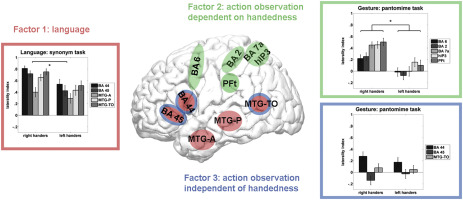
Language, gesture, and handedness are in most people represented in the left cerebral hemisphere. To explore the relations among these attributes, we collected fMRI images in a large sample of left- and right-handers while they performed language tasks and watched action sequences. Regions of interest included the frontal and parietal areas previously identified as comprising an action-observation network, and the frontal and temporal areas comprising the primary areas for language production and comprehension. All of the language areas and most of the action-observation areas showed an overall left-hemispheric bias, despite the participation of equal numbers of left- and right-handers. A factor analysis of the laterality indices derived from the different areas during the tasks indicated three independent networks, one associated with language, one associated with handedness, and one representing action observation independent of handedness. Areas 44 and 45, which together make up Broca's area, were part of the language and action-observation networks, but were not included in the part of the action observation network that was related to handedness, which in turn was strongly linked to areas in the parietal lobe. These results suggest an evolutionary scenario in which the primate mirror neuron system became increasingly lateralized, and later fissioned onto subsystems with one mediating language and the other mediating the execution and observation of manual actions. The second network is further subdivided into one dependent on hand preference and one that is not, providing new insight into the tripartite system of language, handedness, and praxis.
Source:Cortex
Author(s): Isabelle S. Häberling, Paul M. Corballis, Michael C. Corballis
Language, gesture, and handedness are in most people represented in the left cerebral hemisphere. To explore the relations among these attributes, we collected fMRI images in a large sample of left- and right-handers while they performed language tasks and watched action sequences. Regions of interest included the frontal and parietal areas previously identified as comprising an action-observation network, and the frontal and temporal areas comprising the primary areas for language production and comprehension. All of the language areas and most of the action-observation areas showed an overall left-hemispheric bias, despite the participation of equal numbers of left- and right-handers. A factor analysis of the laterality indices derived from the different areas during the tasks indicated three independent networks, one associated with language, one associated with handedness, and one representing action observation independent of handedness. Areas 44 and 45, which together make up Broca's area, were part of the language and action-observation networks, but were not included in the part of the action observation network that was related to handedness, which in turn was strongly linked to areas in the parietal lobe. These results suggest an evolutionary scenario in which the primate mirror neuron system became increasingly lateralized, and later fissioned onto subsystems with one mediating language and the other mediating the execution and observation of manual actions. The second network is further subdivided into one dependent on hand preference and one that is not, providing new insight into the tripartite system of language, handedness, and praxis.
Graphical abstract
Tags: Neurology
ScienceDirect Publication: Brain Research Bulletin
Tropomyosins in the healthy and diseased nervous system
Sunday, June 12, 2016, 16:02
Publication date: Available online 11 June 2016
Source:Brain Research Bulletin
Author(s): Merryn Brettle, Shrujna Patel, Thomas Fath
Regulation of the actin cytoskeleton is dependent on a plethora of actin-associated proteins in all eukaryotic cells. The family of tropomyosins plays a key role in controlling the function of several of these actin-associated proteins and their access to actin filaments. In order to understand the regulation of the actin cytoskeleton in highly dynamic subcellular compartments of neurons such as growth cones of developing neurons and the synaptic compartment of mature neurons, it is pivotal to decipher the functional role of tropomyosins in the nervous system. In this review, we will discuss the current understanding and recent findings on the regulation of the actin cytoskeleton by tropomyosins and potential implication that this has for the dysregulation of the actin cytoskeleton in neurological diseases.
Tags: Neurology
Latest Results for Child's Nervous System
A novel technique to treat acquired Chiari I malformation after supratentorial shunting
Sunday, June 12, 2016, 8:01
Abstract
Purpose
The acquired Chiari I malformation with abnormal cranial vault thickening is a rare late complication of supratentorial shunting. It poses a difficult clinical problem, and there is debate about the optimal surgical strategy. Some authors advocate supratentorial skull enlarging procedures while others prefer a normal Chiari decompression consisting of a suboccipital craniectomy, with or without C1 laminectomy and dural patch grafting.
Methods
We illustrate three cases of symptomatic acquired Chiari I malformation due to inward cranial vault thickening.
Results
We describe a new surgical approach that appears to be effective in these patients. This approach includes the standard Chiari decompression combined with posterior fossa augmentation by thinning the occipital planum.
Conclusion
Internal volume re-expansion of the posterior fossa by thinning the occipital planum appears to be an effective novel surgical strategy in conjunction with the standard surgical therapy of Chiari decompression.
Tags: Neurology
Neuro-Oncology
Antidepressant use and risk of central nervous system metastasis
Sunday, June 12, 2016, 7:26
Abstract
Selective serotonin reuptake inhibitors (SSRIs), a class of antidepressants, were found to increase central nervous system (CNS) metastasis in mice. Our study investigated in humans whether antidepressants, and specifically SSRIs, increased the relative odds of CNS metastasis. We identified 189 cases of CNS metastasis amongst breast cancer, melanoma, and non-Hodgkin lymphoma subjects who were diagnosed with CNS metastasis or infiltration between January 1, 2005 and September 30, 2013 and 756 controls (patients without CNS metastasis or infiltration). Using logistic regression, we estimated the relative odds of CNS metastasis associated with antidepressant use adjusting for relevant covariates. The prevalence of antidepressants was 28.6 % in cases and 27.5 % in controls, whereas SSRIs were used in 16.9 % of cases and 17.3 % of controls. Among all patients, antidepressants were not associated with CNS metastasis or infiltration. No consistent patterns of association were observed in the analyses of other cancer subsets or exposure measures, with the possible exception of an increased risk of CNS metastasis associated with 'any SSRI use' among breast cancer patients (OR = 1.73, 95 % CI = 0.75, 4.04). We did not observe clear patterns of association, which may be due in part to the small sample size in many of our analyses.
Tags: Cancer, Neurology
Behavioural Brain Research
High-speed video gait analysis reveals early and characteristic locomotor phenotypes in mouse models of neurodegenerative movement disorders
Saturday, June 11, 2016, 23:08
Publication date: 15 September 2016
Source:Behavioural Brain Research, Volume 311
Author(s): Daniel F. Preisig, Luka Kulic, Maik Krüger, Fabian Wirth, Jordan McAfoose, Claudia Späni, Pascal Gantenbein, Rebecca Derungs, Roger M. Nitsch, Tobias Welt
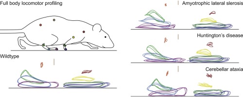
Neurodegenerative diseases of the central nervous system frequently affect the locomotor system resulting in impaired movement and gait. In this study we performed a whole-body high-speed video gait analysis in three different mouse lines of neurodegenerative movement disorders to investigate the motor phenotype. Based on precise computerized motion tracking of all relevant joints and the tail, a custom-developed algorithm generated individual and comprehensive locomotor profiles consisting of 164 spatial and temporal parameters. Gait changes observed in the three models corresponded closely to the classical clinical symptoms described in these disorders: Muscle atrophy due to motor neuron loss in SOD1 G93A transgenic mice led to gait characterized by changes in hind-limb movement and positioning. In contrast, locomotion in huntingtin N171-82Q mice modeling Huntington's disease with basal ganglia damage was defined by hyperkinetic limb movements and rigidity of the trunk. Harlequin mutant mice modeling cerebellar degeneration showed gait instability and extensive changes in limb positioning. Moreover, model specific gait parameters were identified and were shown to be more sensitive than conventional motor tests. Altogether, this technique provides new opportunities to decipher underlying disease mechanisms and test novel therapeutic approaches.
Source:Behavioural Brain Research, Volume 311
Author(s): Daniel F. Preisig, Luka Kulic, Maik Krüger, Fabian Wirth, Jordan McAfoose, Claudia Späni, Pascal Gantenbein, Rebecca Derungs, Roger M. Nitsch, Tobias Welt
Neurodegenerative diseases of the central nervous system frequently affect the locomotor system resulting in impaired movement and gait. In this study we performed a whole-body high-speed video gait analysis in three different mouse lines of neurodegenerative movement disorders to investigate the motor phenotype. Based on precise computerized motion tracking of all relevant joints and the tail, a custom-developed algorithm generated individual and comprehensive locomotor profiles consisting of 164 spatial and temporal parameters. Gait changes observed in the three models corresponded closely to the classical clinical symptoms described in these disorders: Muscle atrophy due to motor neuron loss in SOD1 G93A transgenic mice led to gait characterized by changes in hind-limb movement and positioning. In contrast, locomotion in huntingtin N171-82Q mice modeling Huntington's disease with basal ganglia damage was defined by hyperkinetic limb movements and rigidity of the trunk. Harlequin mutant mice modeling cerebellar degeneration showed gait instability and extensive changes in limb positioning. Moreover, model specific gait parameters were identified and were shown to be more sensitive than conventional motor tests. Altogether, this technique provides new opportunities to decipher underlying disease mechanisms and test novel therapeutic approaches.
Graphical abstract
Tags: Neurology
Journal of NeuroEngineering and Rehabilitation - Latest Articles
A novel robot for imposing perturbations during overground walking: mechanism, control and normative stepping responses
Andrej Olenšek, Matjaž Zadravec And Zlatko Matjačić · Saturday, June 11, 2016, 19:39
The most common approach to studying dynamic balance during walking is by applying perturbations. Previous studies that investigated dynamic balance responses predominantly focused on applying perturbations in...
Tags: Neurology
Current Opinion in Neurobiology
The neurobiology of pair bond formation, bond disruption, and social buffering
Saturday, June 11, 2016, 18:01
Publication date: October 2016
Source:Current Opinion in Neurobiology, Volume 40
Author(s): Claudia Lieberwirth, Zuoxin Wang
Enduring social bonds play an essential role in human society. These bonds positively affect psychological, physiological, and behavioral functions. Here, we review the recent literature on the neurobiology, particularly the role of oxytocin and dopamine, of pair bond formation, bond disruption, and social buffering effects on stress responses, from studies utilizing the socially monogamous prairie vole (Microtus ochrogaster).
Tags: Neurology
Pediatric Neurosurgeons
Pediatric occipitocervical fixation: radiographic criteria, surgical technique, and clinical outcomes based on experience of a single surgeon.
Martinez-del-campo E, Turner Jd, Rangel-castilla L, Soriano-baron H, Kalb S, Theodore N · Saturday, June 11, 2016, 13:20
Pediatric occipitocervical fixation: radiographic criteria, surgical technique, and clinical outcomes based on experience of a single surgeon.
J Neurosurg Pediatr. 2016 Jun 10;:1-11
Authors: Martinez-Del-Campo E, Turner JD, Rangel-Castilla L, Soriano-Baron H, Kalb S, Theodore N
Abstract
OBJECTIVE If left untreated, occipitocervical (OC) instability may lead to serious neurological injury or death. Open internal fixation is often necessary to protect the neurovascular elements. This study reviews the etiologies for pediatric OC instability, analyzes the radiographic criteria for surgical intervention, discusses surgical fixation techniques, and evaluates long-term postoperative outcomes based on a single surgeon's experience. METHODS The charts of all patients < 18 years old who underwent internal OC fixation conducted by the senior author were retrospectively reviewed. Forty consecutive patients were identified for analysis. Patient demographic data, OC junction pathology, radiological diagnostic tools, surgical indications, and outcomes are reported. RESULTS The study population consisted of 20 boys and 20 girls, with a mean age of 7.3 years. Trauma (45% [n = 18]) was the most common cause of instability, followed by congenital etiologies (37.5% [n = 15]). The condyle-C1 interval had a diagnostic sensitivity of 100% for atlantooccipital dislocation. The median number of fixated segments was 5 (occiput-C4). Structural bone grafts were used in all patients. Postsurgical neurological improvement was seen in 88.2% (15/17) of patients with chronic myelopathy and in 25% (1/4) of patients with acute myelopathy. Preoperatively, 42.5% (17/40) of patients were neurologically intact and remained unchanged at last follow-up, 42.5% (17/40) had neurological improvement, 12.5% (5/40) remained unchanged, and 2.5% (1/40) deteriorated. All patients had successful fusion at 1-year follow-up. The complication rate was 7.5% (3/40), including 1 case of vertebral artery injury. CONCLUSIONS Occipitocervical fixation is safe in children and provides immediate immobilization, with excellent survival and arthrodesis rates. Of the radiographic tools evaluated, the condyle-C1 interval was the most predictive of atlantooccipital dislocation.
OBJECTIVE If left untreated, occipitocervical (OC) instability may lead to serious neurological injury or death. Open internal fixation is often necessary to protect the neurovascular elements. This study reviews the etiologies for pediatric OC instability, analyzes the radiographic criteria for surgical intervention, discusses surgical fixation techniques, and evaluates long-term postoperative outcomes based on a single surgeon's experience. METHODS The charts of all patients < 18 years old who underwent internal OC fixation conducted by the senior author were retrospectively reviewed. Forty consecutive patients were identified for analysis. Patient demographic data, OC junction pathology, radiological diagnostic tools, surgical indications, and outcomes are reported. RESULTS The study population consisted of 20 boys and 20 girls, with a mean age of 7.3 years. Trauma (45% [n = 18]) was the most common cause of instability, followed by congenital etiologies (37.5% [n = 15]). The condyle-C1 interval had a diagnostic sensitivity of 100% for atlantooccipital dislocation. The median number of fixated segments was 5 (occiput-C4). Structural bone grafts were used in all patients. Postsurgical neurological improvement was seen in 88.2% (15/17) of patients with chronic myelopathy and in 25% (1/4) of patients with acute myelopathy. Preoperatively, 42.5% (17/40) of patients were neurologically intact and remained unchanged at last follow-up, 42.5% (17/40) had neurological improvement, 12.5% (5/40) remained unchanged, and 2.5% (1/40) deteriorated. All patients had successful fusion at 1-year follow-up. The complication rate was 7.5% (3/40), including 1 case of vertebral artery injury. CONCLUSIONS Occipitocervical fixation is safe in children and provides immediate immobilization, with excellent survival and arthrodesis rates. Of the radiographic tools evaluated, the condyle-C1 interval was the most predictive of atlantooccipital dislocation.
PMID: 27286444 [PubMed - as supplied by publisher]
Tags: Neurology
Pediatric Neurosurgeons
Physiological growth hormone replacement and rate of recurrence of craniopharyngioma: the Genentech National Cooperative Growth Study.
Smith Tr, Cote Dj, Jane Ja, Laws Er · Saturday, June 11, 2016, 13:20
Physiological growth hormone replacement and rate of recurrence of craniopharyngioma: the Genentech National Cooperative Growth Study.
J Neurosurg Pediatr. 2016 Jun 10;:1-5
Authors: Smith TR, Cote DJ, Jane JA, Laws ER
Abstract
OBJECTIVE The object of this study was to establish recurrence rates in patients with craniopharyngioma postoperatively treated with recombinant human growth hormone (rhGH) as a basis for determining the risk of rhGH therapy in the development of recurrent tumor. METHODS The study included 739 pediatric patients with craniopharyngioma who were naïve to GH upon entering the Genentech National Cooperative Growth Study (NCGS) for treatment. Reoperation for tumor recurrence was documented as an adverse event. Cox proportional-hazards regression models were developed for time to recurrence, using age as the outcome and enrollment date as the predictor. Patients without recurrence were treated as censored. Multivariate logistic regression was used to examine the incidence of recurrence with adjustment for the amount of time at risk. RESULTS Fifty recurrences in these 739 surgically treated patients were recorded. The overall craniopharyngioma recurrence rate in the NCGS was 6.8%, with a median follow-up time of 4.3 years (range 0.7-6.4 years.). Age at the time of study enrollment was statistically significant according to both Cox (p = 0.0032) and logistic (p < 0.001) models, with patients under 9 years of age more likely to suffer recurrence (30 patients [11.8%], 0.025 recurrences/yr of observation, p = 0.0097) than those ages 9-13 years (17 patients [6.0%], 0.17 recurrences/yr of observation) and children older than 13 years (3 patients [1.5%], 0.005 recurrences/yr of observation). CONCLUSIONS Physiological doses of GH do not appear to increase the recurrence rate of craniopharyngioma after surgery in children, but long-term follow-up of GH-treated patients is required to establish a true natural history in the GH treatment era.
OBJECTIVE The object of this study was to establish recurrence rates in patients with craniopharyngioma postoperatively treated with recombinant human growth hormone (rhGH) as a basis for determining the risk of rhGH therapy in the development of recurrent tumor. METHODS The study included 739 pediatric patients with craniopharyngioma who were naïve to GH upon entering the Genentech National Cooperative Growth Study (NCGS) for treatment. Reoperation for tumor recurrence was documented as an adverse event. Cox proportional-hazards regression models were developed for time to recurrence, using age as the outcome and enrollment date as the predictor. Patients without recurrence were treated as censored. Multivariate logistic regression was used to examine the incidence of recurrence with adjustment for the amount of time at risk. RESULTS Fifty recurrences in these 739 surgically treated patients were recorded. The overall craniopharyngioma recurrence rate in the NCGS was 6.8%, with a median follow-up time of 4.3 years (range 0.7-6.4 years.). Age at the time of study enrollment was statistically significant according to both Cox (p = 0.0032) and logistic (p < 0.001) models, with patients under 9 years of age more likely to suffer recurrence (30 patients [11.8%], 0.025 recurrences/yr of observation, p = 0.0097) than those ages 9-13 years (17 patients [6.0%], 0.17 recurrences/yr of observation) and children older than 13 years (3 patients [1.5%], 0.005 recurrences/yr of observation). CONCLUSIONS Physiological doses of GH do not appear to increase the recurrence rate of craniopharyngioma after surgery in children, but long-term follow-up of GH-treated patients is required to establish a true natural history in the GH treatment era.
PMID: 27286443 [PubMed - as supplied by publisher]
Tags: Neurology
Journal of Neuroscience Methods
Anatomy and surgical approach of rat's vestibular sensors and nerves
Saturday, June 11, 2016, 11:54
Publication date: 1 September 2016
Source:Journal of Neuroscience Methods, Volume 270
Author(s): Martin Hitier, Go Sato, Yan-feng Zhang, Yiwen Zheng, Stephane Besnard, Paul F. Smith, Ian S. Curthoys
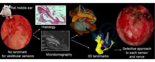
Background The rat is one of the most used species in the neurosciences, but how to selectively reach each of its 5 vestibular sensors has never been described. Besides, new functions of the vestibular system have been recently discovered in the rat involving vegetative, circadian and cognitive functions. But the central pathways sustaining these functions and the role of each of the vestibular sensors are not clear. New methods Here we want to describe the anatomy and look for a direct surgical approach to the 5 vestibular sensors in rats, as an indispensable technique to further study the central vestibular pathways. To do so we studied 10 rats either by microtomography with osmium tetroxide staining, histology with hematoxilyn-eosine staining or microsurgical dissection. Results The microtomography allows a 3D representation of the 5 vestibular sensors and their nerves, with precise landmarks confirmed by the histological analysis. Each of the landmarks are illustrated and a selective surgical approach to each sensor and their nerves, is described step by step. Comparison with existing method Selective approaches to the vestibular sensors have been used in other species such as cats, monkeys and recently humans but the current study is the first allowing this technique in rats. Conclusion Each vestibular sensor of the rat can be reached by a selective surgical approach. This allows further techniques such as electrophysiology or neurotracing of the central vestibular pathways. This also indicates the rat as a potential model for vestibular prostheses.
Source:Journal of Neuroscience Methods, Volume 270
Author(s): Martin Hitier, Go Sato, Yan-feng Zhang, Yiwen Zheng, Stephane Besnard, Paul F. Smith, Ian S. Curthoys
Background The rat is one of the most used species in the neurosciences, but how to selectively reach each of its 5 vestibular sensors has never been described. Besides, new functions of the vestibular system have been recently discovered in the rat involving vegetative, circadian and cognitive functions. But the central pathways sustaining these functions and the role of each of the vestibular sensors are not clear. New methods Here we want to describe the anatomy and look for a direct surgical approach to the 5 vestibular sensors in rats, as an indispensable technique to further study the central vestibular pathways. To do so we studied 10 rats either by microtomography with osmium tetroxide staining, histology with hematoxilyn-eosine staining or microsurgical dissection. Results The microtomography allows a 3D representation of the 5 vestibular sensors and their nerves, with precise landmarks confirmed by the histological analysis. Each of the landmarks are illustrated and a selective surgical approach to each sensor and their nerves, is described step by step. Comparison with existing method Selective approaches to the vestibular sensors have been used in other species such as cats, monkeys and recently humans but the current study is the first allowing this technique in rats. Conclusion Each vestibular sensor of the rat can be reached by a selective surgical approach. This allows further techniques such as electrophysiology or neurotracing of the central vestibular pathways. This also indicates the rat as a potential model for vestibular prostheses.
Graphical abstract
Tags: Neurology
North American Neuro-Ophthalmology Society
Maternally Inherited Diabetes and Deafness is Phenotypically and Genotypically Heterogeneous.
Finsterer, Josef; Frank, Marlies · Saturday, June 11, 2016, 11:37
Tags: Neurology
North American Neuro-Ophthalmology Society
Maternally Inherited Diabetes and Deafness Is Phenotypically and Genotypically Heterogeneous: A Response.
Kattah, Jorge C.; Cardenas, Simon R. · Saturday, June 11, 2016, 11:37
Tags: Neurology
NeuroReport - Published Ahead-of-Print
Transcranial direct current stimulation modulates pattern separation.
Cappiello, Marcus; Xie, Weizhen; David, Alexander; Bikson, Marom; Zhang, Weiwei ·Saturday, June 11, 2016, 11:04
Maintaining similar memories in a distinct and nonoverlapping manner, known as pattern separation, is an important mnemonic process. The medial temporal lobe, especially the hippocampus, has been implicated in this crucial memory function. The present study thus examines whether it is possible to modulate pattern separation using bilateral transcranial direct current stimulation (tDCS) over the temporal lobes. Specifically, in this study, pattern separation was assessed using the Mnemonic Similarity Task following 15-min offline bilateral temporal lobe tDCS (left cathode and right anode or left anode and right cathode) or sham stimulation. In the Mnemonic Similarity Task, participants studied a series of sequentially presented visual objects. In the subsequent recognition memory test, participants viewed a series of sequentially presented objects that could be old images from study, novel foils, or lures that were visually similar to the studied images. Participants reported whether these images were exactly the same as, similar to, or different from the studied images. Following both active tDCS conditions, participants were less likely to identify lures as 'similar' compared with the sham condition, indicating a reduction in pattern separation resulting from temporal lobe tDCS. In contrast, no significant difference in overall accuracy was found for participants' discrimination of old and new images. Together, these results suggest that temporal lobe tDCS can selectively modulate the pattern separation function without changing participants' baseline recognition memory performance. (C) 2016 Wolters Kluwer Health | Lippincott Williams & Wilkins
Tags: Neurology
European Journal of Neuroscience
Rizatriptan overuse promotes hyperalgesia induced by dural inflammatory stimulation in rats by modulation of the serotonin system
Min Su, Ye Ran, Xun Han, Yufei Liu, Xu Zhang, Qingche Tan, Ruisheng Li, Shengyuan Yu · Saturday, June 11, 2016, 10:06
Abstract
Clinical and preclinical studies have implicated serotonin (5-HT) and the 5-HT2A receptor (5-HT2AR) in the pathogenesis of medication-overuse headache (MOH). However, with no appropriate animal model to study this phenomenon it is difficult to differentiate the effects of chronic exposure to analgesics from the consequences of frequent headache attacks during the development of MOH. Therefore, this study used a novel animal model of MOH established by a combination of the overuse of rizatriptan (RIZ) and stimulation with dural inflammatory soup (IS) to investigate whether 5-HT and 5-HT2AR are involved in central plasticity and hyperalgesia. Similar to an IS infusion, IS-RIZ treatment induced nociception-related behaviours in Sprague-Dawley rats and increased Fos expression in the cortex and trigeminal pathway whereas the RIZ injection alone did not. Additionally, overuse of RIZ, administration of an IS stimulus, and a combination of these treatments, decreased the pentobarbital withdrawal threshold, with IS-RIZ treatment having the most significant effects. Both chronic RIZ exposure and recurring nociception decreased 5-HT expression whereas IS-RIZ treatment led to decreased expression of 5-HT and upregulation of 5-HT2AR, which was positively correlated with Fos activation. These findings suggest that overuse of RIZ does not directly induce pain via the activation of nociceptive pathways but may increase hyperalgesia by influencing the pain modulation system. Furthermore, decreased 5-HT levels and upregulation of 5-HT2AR may play important roles in this system. Taken together, these findings indicate that medication overuse and frequent headache attacks can promote the neural plasticity associated with MOH.
This article is protected by copyright. All rights reserved.
Tags: Neurology
Journal of Neurotrauma
Screening for Post-Traumatic Stress Disorder in a Civilian Emergency Department Population with Traumatic Brain Injury
Juliet Haarbauer-krupa · Saturday, June 11, 2016, 8:59
Journal of Neurotrauma , Vol. 0, No. 0.
Tags: Neurology
Journal of Neurotrauma
Human Mesenchymal Stem Cell Treatment Normalizes Cortical Gene Expression after Traumatic Brain Injury
Ali Darkazalli · Saturday, June 11, 2016, 8:59
Journal of Neurotrauma , Vol. 0, No. 0.
Tags: Neurology
Journal of Neurotrauma
Brain Volume, Connectivity, and Neuropsychological Performance in Mild Traumatic Brain Injury: The Impact of Post-Traumatic Stress Disorder Symptoms
Katherine C. Lopez · Saturday, June 11, 2016, 8:59
Journal of Neurotrauma , Vol. 0, No. 0.
Tags: Neurology
Journal of Neurotrauma
Trauma-Specific Brain Abnormalities in Suspected Mild Traumatic Brain Injury Patients Identified in the First 48 Hours after Injury: A Blinded Magnetic Resonance Imaging Comparative Study Including Suspected Acute Minor Stroke Patients
Maria Chiara Ricciardi · Saturday, June 11, 2016, 8:59
Journal of Neurotrauma , Vol. 0, No. 0.
Tags: Neurology
Journal of Neurotrauma
Pioglitazone Attenuates Neuroinflammation and Promotes Dopaminergic Neuronal Survival in the Nigrostriatal System of Rats after Diffuse Brain Injury
Mei Liu · Saturday, June 11, 2016, 8:59
Journal of Neurotrauma , Vol. 0, No. 0.
Tags: Neurology
Journal of Neurotrauma
Influence of Previous Comorbidities and Common Complications on Motor Function after Early Surgical Treatment of Patients with Traumatic Spinal Cord Injury
Michael Kreinest · Saturday, June 11, 2016, 8:59
Journal of Neurotrauma , Vol. 0, No. 0.
Tags: Neurology
Journal of Neurotrauma
Blood Aggravates Histological and Functional Damage after Acute Subdural Hematoma in Rats
Daniel Jussen · Saturday, June 11, 2016, 8:59
Journal of Neurotrauma , Vol. 0, No. 0.
Tags: Neurology
Journal of Neurotrauma
On the Limitations of Progesterone Treatment in Very Severe Traumatic Brain Injury: What Can Be Learned from Allitt et al., "Progesterone Exacerbates Short-Term Effects of Traumatic Brain Injury"
Fahim Atif · Saturday, June 11, 2016, 8:59
Journal of Neurotrauma , Vol. 0, No. 0.
Tags: Neurology
Latest Results for Child's Nervous System
Anatomy of spinal nerves in the first Turkish illustrated anatomy handwritten textbook
Saturday, June 11, 2016, 8:00
Tags: Neurology
Neuro-Oncology
Single-agent erlotinib versus oral etoposide in patients with recurrent or refractory pediatric ependymoma: a randomized open-label study
Saturday, June 11, 2016, 7:25
Abstract
Overexpression of human epidermal growth factor receptor (HER/EGFR) is associated with various tumors, including ependymomas. To investigate whether EGFR inhibition was of benefit in pediatric patients with recurrent ependymoma, a multi-center, randomized, open-label, phase 2 study of oral erlotinib versus oral etoposide was undertaken. Twenty-five patients were randomized to receive erlotinib 85 mg/m2 daily or etoposide 50 mg/m2/day for 21 consecutive days followed by a 7-day rest period. Courses were repeated every 28 days. In the erlotinib arm, no patient achieved a complete, partial, or minor response, and only 2 (15.4 %) patients showed stable disease as their best response. In the etoposide arm, 2 patients (16.7 %) demonstrated partial responses, 1 (8.3 %) patient demonstrated a minor response, and 2 (16.7 %) showed prolonged stable disease, for a prolonged disease control rate of 41.7 %. Three patients received at least nine cycles of etoposide (range 9–24 cycles) before discontinuing at the request of the physician and/or family. Four patients who failed etoposide in this study received erlotinib in a companion single arm study; none had a response. The futility criteria were met at the second interim analysis, and both studies were discontinued. Pharmacokinetics of erlotinib were similar to previous observations in pediatric patients. Overall, erlotinib was well tolerated and safety was consistent with its established profile in adults. The overall risk–benefit profile does not support the use of erlotinib in pediatric patients with recurrent ependymoma, whereas single-agent etoposide appears to have efficacy in a subset of patients.
Tags: Cancer, Neurology
World Neurosurgery
Outcome of Endoscopic Third Ventriculostomy in Pediatric Patients at Zewditu Memorial Hospital, Ethiopia
Saturday, June 11, 2016, 4:59
Publication date: August 2016
Source:World Neurosurgery, Volume 92
Author(s): Hagos Biluts, Azarias Kassahun Admasu
Objective To determine short-term outcome of endoscopic third ventriculostomy (ETV) in pediatric patients. Methods This was a hospital-based retrospective study of outcome of ETV performed with or without choroid plexus cauterization (CPC) in pediatric patients at Zewditu Memorial Hospital, Addis Ababa, Ethiopia, between January 2012 and December 2014. Medical records were used to complete a structured questionnaire. Outcomes were graded as success or failure. The difference in proportions was examined using χ2test. Results Of 122 children, 26 underwent a combined ETV and CPC procedure, and 96 underwent ETV alone. The mean and median ages were 1.89 months and 2.0 months, respectively. Most patients were <6 months old (45.1%); 35.2% were >1 year old. The cause of hydrocephalus was myelomeningocele in 42 (34.4%) patients and aqueductal stenosis in 41 (33.6%) patients. The mean and median follow-up periods were 7.2 months and 3.0 months, respectively. The success rate for combined ETV and CPC (66%) was superior to the success rate for ETV alone (47%) among infants <1 year old (P < 0.0001). The overall surgical mortality rate was 2.4%, and the infection rate was 7%. Conclusions In resource-limited countries, ETV can be performed with acceptable results and less mortality and morbidity. Myelomeningocele and aqueductal stenosis were the most common causes of hydrocephalus. In patients <1 year old, the outcome success in patients undergoing combined ETV and CPC (53%) was significantly better compared with patients undergoing ETV alone (25%).
Tags: Neurology
World Neurosurgery
Seizure Correlates with Prolonged Hospital Stay, Increased Costs, and Increased Mortality in Nontraumatic Subdural Hematoma
Saturday, June 11, 2016, 4:59
Publication date: August 2016
Source:World Neurosurgery, Volume 92
Author(s): Jacob R. Joseph, Brandon W. Smith, Craig A. Williamson, Paul Park
Background Nontraumatic subdural hematoma (NTSDH) is a common neurosurgical disease process, with mortality reported as high as 13%. Seizure has a known association with NTSDH, although patient outcomes have not previously been well studied in this population. The purpose of this study was to examine the relationship between in-hospital seizure and inpatient outcomes in NTSDH. Methods Using the University HealthSystem Consortium (UHC) database, we performed a retrospective cohort study of adults with a principal diagnosis of NTSDH (International Classification of Diseases, Ninth Revision code 43.21) between 2011 and 2015. Patients with in-hospital seizure (International Classification of Diseases, Ninth Revision codes 34500–34591, 78033, 78039) were compared with those without. Patients with a history of seizure before arrival were excluded. Patient demographics, hospital length of stay (LOS), intensive care unit stay, in-hospital mortality, and direct costs were recorded. Results A total 16,928 patients with NTSDH were identified. Mean age was 69.2 years, and 64.7% were male. In-hospital seizure was documented in 744 (4.40%) patients. Hospital LOS was 17.64 days in patients with seizure and 6.26 days in those without (P < 0.0001). Mean intensive care unit stay increased from 3.36 days without seizure to 9.36 days with seizure. In-hospital mortality was 9.19% in patients without seizure and 16.13% in those with seizure (P < 0.0001). Direct costs were $12,781 in patients without seizure and $38,110 in those with seizure (P < 0.0001). Conclusions Seizure in patients with NTSDH correlates with significantly increased total LOS and increased mortality. Direct costs are similarly increased. Further studies accounting for effects of illness severity are necessary to validate these results.
Tags: Neurology
World Neurosurgery
Smith-Robinson Procedure with an Autologous Iliac Crest for Degenerative Cervical Disc Disease: A 28-Year Follow-Up of 95 Patients
Saturday, June 11, 2016, 4:59
Publication date: August 2016
Source:World Neurosurgery, Volume 92
Author(s): Benedikt W. Burkhardt, Moritz Brielmaier, Karsten Schwerdtfeger, Salam Sharif, Joachim M. Oertel
Background Long-term clinical results after anterior cervical discectomy and fusion (ACDF) with an autologous iliac crest are rare. The purpose of this study was to assess this, with special focus on pain, functional outcome, and repeat surgery for adjacent segment disease (ASD). Methods Hospital records of 212 patients who were affected by degenerative cervical disc disease and treated by the Smith-Robinson technique were reviewed. Information about diagnosis, surgery, pre- and postoperative clinical process, and complications was analyzed. Patients were reviewed with a standardized questionnaire including the current neurologic status, Neck Disability Index, EQ-5D, Patient Satisfaction Index, Odom criteria, and limitations in quality of life. Results Ninety-five patients with a mean follow-up of 28 years were evaluated. ACDF was performed at 1 level in 67 and 2 levels in 28 patients. Ninety-two patients reported pain before surgery and 68 patients remained pain free and did not require second surgery. At follow-up, the mean Neck Disability Index was 14%, and mean EQ-5D score was 5. Postoperatively, 96.8% of patients were satisfied and 84.2% of patients reported good to excellent functional recovery. One patient had a hairline fracture at the iliac crest donor site. Fourteen patients underwent second surgery because of degenerative changes, including 11 at the symptomatic ASD. Conclusions ACDF yields significant decrease in pain, a significant increase in function, and a high degree of patient satisfaction. Overall prevalence for ASD was 12.0% after 25 years. Patients with reoperation had similar clinical outcome regarding pain, compared with patients without reoperation.
Tags: Neurology
World Neurosurgery
Impact of Neurosurgery Medical Student Research Grants on Neurosurgery Residency Choice
Saturday, June 11, 2016, 4:59
Publication date: August 2016
Source:World Neurosurgery, Volume 92
Author(s): Ahmed J. Awad, Christopher A. Sarkiss, Christopher P. Kellner, Jeremy Steinberger, Justin R. Mascitelli, Eric K. Oermann, Margaret Pain, Reade De Leacy, Raj Shrivastava, Joshua B. Bederson, J Mocco
Background Recent decades have seen a rapid expansion of involvement of medical students in biomedical research during medical school training. Research within medical school has been shown to influence medical students with regard to medical knowledge, career development, and residency specialty choice. The objective of this study was to evaluate the impact of neurosurgery medical student research grants on neurosurgery residency choice and provide an insight on the demographics of grant awardees. Methods In this retrospective study, a search of award recipients was performed using data available on the American Association of Neurological Surgeons, Congress of Neurological Surgeons, and Neurosurgery Research and Education Foundation websites. Searched years included the first cycle of American Association of Neurological Surgeons/Neurosurgery Research and Education Foundation (2007) and Council of State Neurosurgical Societies/Congress of Neurological Surgeons (2008–2009) grant awards until the 2015–2016 cycle, which is the latest award cycle to date. Results The initial search yielded 163 research grants that were awarded to 158 students between the years of 2007 and 2016. Among the 163 grant recipients, 126 (77.3%) were men. Among the 88 recipients who entered postgraduate residency programs, 51% (45 of 88) matched into neurosurgery residency. When considering both neurosurgery and neurology residency programs, the percentage increased to 59.1% (52 of 88). Conclusions Neurosurgery grants for medical students are highly successful in producing future neurosurgeons with >50% of grant recipients matched into neurosurgery. Women are underrepresented in neurosurgery grants and neurosurgery residency programs. This situation can be improved by providing insight about the field early in medical school, perhaps through increased use of neurosurgery medical student grants.
Tags: Neurology
Behavioural Brain Research
Sustained lentiviral-mediated overexpression of microRNA124a in the dentate gyrus exacerbates anxiety- and autism-like behaviors associated with neonatal isolation in rats
Friday, June 10, 2016, 23:07
Publication date: 15 September 2016
Source:Behavioural Brain Research, Volume 311
Author(s): Amine Bahi
Autism spectrum disorders (ASD) are highly disabling psychiatric disorders. Despite a strong genetic etiology, there are no efficient therapeutic interventions that target the core symptoms of ASD. Emerging evidence suggests that dysfunction of microRNA (miR) machinery may contribute to the underlying molecular mechanisms involved in ASD. Here, we report a stress model demonstrating that neonatal isolation-induced long-lasting hippocampal elevation of miR124a was associated with reduced expression of its target BDNF mRNA. In addition, we investigated the impact of lentiviral-mediated overexpression of miR124a into the dentate gyrus (DG) on social interaction, repetitive- and anxiety-like behaviors in the neonatal isolation (Iso) model of autism. Rats isolated from the dams on PND 1 to PND 11 were assessed for their social interaction, marble burying test (MBT) and repetitive self-grooming behaviors as adults following miR124a overexpression. Also, anxiety-like behavior and locomotion were evaluated in the elevated plus maze (EPM) and open-field (OF) tests. Results show that, consistent with previously published reports, Iso rats displayed decreased social interaction contacts but increased repetitive- and anxiety-like behaviors. Interestingly, across both autism- and anxiety-like behavioral assays, miR124a overexpression in the DG significantly exacerbated repetitive behaviors, social impairments and anxiety with no effect on locomotor activity. Our novel findings attribute neonatal isolation-inducible cognitive impairments to induction of miR124a and consequently suppressed BDNF mRNA, opening venues for intercepting these miR124a-mediated damages. They also highlight the importance of studying microRNAs in the context of ASD and identify miR124a as a novel potential therapeutic target for improving mood disorders.
Tags: Neurology
Behavioural Brain Research
The flavonoid baicalein rescues synaptic plasticity and memory deficits in a mouse model of Alzheimer's disease
Friday, June 10, 2016, 23:07
Publication date: 15 September 2016
Source:Behavioural Brain Research, Volume 311
Author(s): Xun-Hu Gu, Li-Jun Xu, Zhi-Qiang Liu, Bo Wei, Yuan-Jian Yang, Guo-Gang Xu, Xiao-Ping Yin, Wei Wang
Increasing evidence suggests that disruptions of synaptic functions correlate with the severity of cognitive deficit in Alzheimer's disease (AD). Our previous study demonstrated that baicalein enhances long-term potentiation (LTP) in acute rat hippocampal slices and improves hippocampus-dependent contextual fear conditioning in rats. Given that baicalein possess various biological activities, especially its effects on synaptic plasticity and cognitive function, we examined the effect of baicalein on synaptic function both in vitro and in vivo in AD model. The effect of baicalein on Aβ42 oligomer impaired LTP was investigated by electrophysiological methods. Baicalein was administered orally via drinking water to the APP/PS1 mice and sex- and age-matched wild-type mice. Treatment started at 5 months of age and mice were assessed for cognition and AD-like pathology at 7-month-old. Cognition was analyzed by Morris water maze test, fear conditioning test, and novel object recognition test. Changes in hippocampal 12/15 Lipoxygenase (12/15LO) and glycogen synthase kinase 3β (GSK3β) activity, Aβ production, tau phosphorylation, synaptic plasticity, and dendritic spine density were evaluated. Baicalein prevented Aβ-induced impairments in hippocampal LTP through activation of serine threonine Kinase (Akt) phosphorylation. Long-term oral administration of baicalein inhibited 12/15LO and GSK3β activity, reduced β-secretase enzyme (BACE1), decreased the concentration of total Aβ, and prevented phosphorylation of tau in APP/PS1 mice. Meanwhile, baicalein restored spine number, synaptic plasticity, and memory deficits. Our results strengthen the potential of the flavonoid baicalein as a novel and promising oral bioactive therapeutic agent that prevents memory deficits in AD.
Tags: Neurology
Behavioural Brain Research
Time-dependent effects of repeated THC treatment on dopamine D2/3 receptor-mediated signalling in midbrain and striatum
Friday, June 10, 2016, 23:07
Publication date: 15 September 2016
Source:Behavioural Brain Research, Volume 311
Author(s): Benjamin B. Tournier, Stergios Tsartsalis, Andrea Dimiziani, Philippe Millet, Nathalie Ginovart
This study examined the time-course of alterations in levels and functional sensitivities of dopamine D2/3 receptors (D2/3R) during the course and up to 6 weeks following cessation of chronic treatment with Delta(9)-Tetrahydrocannabinol (THC) in rats. THC treatment led to an increase in D2/3R levels in striatum, as assessed using [3H]-(+)-PHNO, that was readily observable after one week of treatment, remained stably elevated during the subsequent 2 weeks of treatment, but fully reversed within 2 weeks of THC discontinuation. THC-induced D2/3R alterations were more pronounced and longer lasting in the dopamine cell body regions of the midbrain, wherein [3H]-(+)-PHNO binding was still elevated at 2 weeks but back to control values at 6 weeks after THC cessation. Parallel analyses of the psychomotor effects of pre- and post-synaptic doses of quinpirole also showed a pattern of D2/3R functional supersensitivity indicative of more rapid subsidence in striatum than in midbrain following drug cessation. These results indicate that chronic THC is associated with a biochemical and functional sensitization of D2/3R signaling, that these responses show a region-specific temporal pattern and are fully reversible following drug discontinuation. These results suggest that an increased post-synaptic D2/3R function and a decreased DA presynaptic signaling, mediated by increased D2/3R autoinhibition, may predominate during distinct phases of withdrawal and may contribute both to the mechanisms leading to relapse and to cannabinoid withdrawal symptoms. The different rates of normalization of D2/3R function in striatum and midbrain may be critical information for the development of new pharmacotherapies for cannabis dependence.
Tags: Neurology
Behavioural Brain Research
Quantitative proteomics analysis of the liver reveals immune regulation and lipid metabolism dysregulation in a mouse model of depression
Friday, June 10, 2016, 23:07
Publication date: 15 September 2016
Source:Behavioural Brain Research, Volume 311
Author(s): You Wu, Jianyong Tang, Chanjuan Zhou, Libo Zhao, Jin Chen, Li Zeng, Chenglong Rao, Haiyang Shi, Li Liao, Zihong Liang, Yongtao Yang, Jian Zhou, Peng Xie
Major depressive disorder (MDD) is a highly prevalent and debilitating mental illness with substantial impairments in quality of life and functioning. However, the pathophysiology of major depression remains poorly understood. Combining the brain and body should provide a comprehensive understanding of the etiology of MDD. As the largest internal organ of the human body, the liver has an important function, yet no proteomic study has assessed liver protein expression in a preclinical model of depression. Using the chronic unpredictable mild stress (CUMS) mouse model of depression, differential protein expression between CUMS and control (CON) mice was examined in the liver proteome using isobaric tag for relative and absolute quantitation (iTRAQ) coupled with tandem mass spectrometry. More than 4000 proteins were identified and 66 most significantly differentiated proteins were used for further bioinformatic analysis. According to the ingenuity pathway analysis (IPA), we found that proteins related to the inflammation response, immune regulation, lipid metabolism and NFκB signaling network were altered by CUMS. Moreover, four proteins closely associated with these processes, hemopexin, haptoglobin, cytochrome P450 2A4 (CYP2A4) and bile salt sulfotransferase 1 (SULT2A1), were validated by western blotting. In conclusion, we report, for the first time, the liver protein expression profile in the CUMS mouse model of depression. Our findings provide novel insight (liver–brain axis) into the multifaceted mechanisms of major depressive disorder.
Tags: Neurology
Cortex
Reading words and other people: a comparison of exception word, familiar face and affect processing in the left and right temporal variants of primary progressive aphasia
Friday, June 10, 2016, 20:18
Publication date: Available online 9 June 2016
Source:Cortex
Author(s): Richard J. Binney, Maya Henry, Miranda Babiak, Peter S. Pressman, Miguel A. Santos, Jared Narvid, Maria Luisa Mandelli, Paul J. Strain, Bruce L. Miller, Katherine P. Rankin, Howard J. Rosen, Maria Luisa Gorno-Tempini
Semantic variant primary progressive aphasia (svPPA) typically presents with left-hemisphere predominant rostral temporal lobe atrophy and the most significant complaints within the language domain. Less frequently, patients present with right-hemisphere predominant temporal atrophy coupled with marked impairments in processing of famous faces and emotions. Few studies have objectively compared these patient groups in both domains and therefore it is unclear to what extent the syndromes overlap. Clinically diagnosed svPPA patients were characterized as left- (n= 21) or right-predominant (n = 12) using imaging and compared along with 14 healthy controls. Regarding language, our primary focus was upon two hallmark features of svPPA; confrontation naming and surface dyslexia. Both groups exhibited naming deficits and surface dyslexia although the impairments were more severe in the left-predominant group. Familiarity judgments on famous faces and affect processing were more profoundly impaired in the right-predominant group. Our findings suggest that the two syndromes overlap significantly but that early cases at the tail ends of the continuum constitute a challenge for current clinical criteria. Correlational neuroimaging analyses implicated a mid portion of the left lateral temporal lobe in exception word reading impairments in line with proposals that this region is an interface between phonology and semantic knowledge.
Tags: Neurology
Journal of NeuroEngineering and Rehabilitation - Latest Articles
Impact on gait biomechanics of using an active variable impedance prosthetic knee
Matthew R. Williams, Susan D'andrea And Hugh M. Herr · Friday, June 10, 2016, 19:34
An above knee amputation can have a significant impact on gait, with substantial deviations in inter-leg symmetry, step length, hip exertion and upper body involvement even when using a current clinical standa...
Tags: Neurology
European Journal of Neuroscience
Peripheral and central alterations affecting spinal nociceptive processing and pain at adulthood in rats exposed to neonatal maternal deprivation
Pierre-eric Juif, Chiara Salio, Vivien Zell, Meggane Melchior, Adrien Lacaud, Nathalie Petit-demouliere, Francesco Ferrini, Pascal Darbon, Ulrike Hanesch, Fernand Anton, Adalberto Merighi, Vincent Lelièvre, Pierrick Poisbeau · Friday, June 10, 2016, 17:58
Abstract
The nociceptive system of rodents is not fully developed and functional at birth. Specifically, C fibers transmitting peripheral nociceptive information establish synaptic connections in the spinal cord already during the embryonic period that only become fully functional after birth. Here, we studied the consequences of neonatal maternal deprivation (NMD, 3h/day, P2-P12) on the functional establishment of C fiber-mediated neurotransmission in spinal cord and of pain-related behavior. In vivo recording revealed that C fiber-mediated excitation of spinal cord neurons could be observed at P14 only in control but not in NMD rats. NMD was associated with a strong alteration in the expression of growth factors controlling C nociceptor maturation as well as K2P channels known to set nociceptive thresholds. In good agreement, C-type sensory neurons from NMD animals appeared to be hypoexcitable but functionally connected to spinal neurons, especially those expressing TRPV1 receptors. In vivo and in vitro recordings of lamina II spinal neurons at P14 revealed that the NMD-related lack of C fiber-evoked responses resulted from an inhibitory barrage in the spinal cord dorsal horn. Eventually, C-type sensory-spinal processing could be recovered after a delay of about ten days in NMD animals. However, animals remained hypersensitive to noxious stimulus up to P100 and this might be due to an excessive expression of Nav1.8 transcripts in DRG neurons. Together, our data provide evidence for a deleterious impact of perinatal stress exposure on the maturation of the sensory-spinal nociceptive system that may contribute to the nociceptive hypersensitivity in early adulthood.
This article is protected by copyright. All rights reserved.
Tags: Neurology
European Journal of Neuroscience
Brain-derived neurotrophic factor prevents dendritic retraction of adult mouse retinal ganglion cells
Kate E Binley, Wai S Ng, Yves-alain Barde, Bing Song, James E Morgan · Friday, June 10, 2016, 17:58
Abstract
We used cultured adult mouse retinae as a model system to follow and quantify the retraction of dendrites using diolistic labelling of retinal ganglion cells (RGCs) following explantation. Cell death was monitored in parallel by nuclear staining as staining withRGC and apoptotic markers was inconsistent and exceedingly difficult to reliably quantify. Nuclear staining allowed us to delineate a lengthy time window during which dendrite retraction can be monitored in the absence of RGC death. The addition of brain-derived neurotrophic factor (BDNF) produced a marked reduction in dendritic degeneration, even when application was delayed for 3 days after retina explantation. These results suggest that the delayed addition of trophic factors may be functionally beneficial before the loss of cell bodies in the course of conditions such as glaucoma.
This article is protected by copyright. All rights reserved.
Tags: Neurology
European Journal of Neuroscience
Present-self, past-self and the close-other: Neural correlates of assigning trait adjectives to oneself and others
Ilona Kotlewska, Anna Nowicka · Friday, June 10, 2016, 17:58
Abstract
Information regarding the past-self may be viewed as information referring to other people. However, evidence supporting this notion at the neural level is rather sparse and it remains unclear whether the past-self is processed like any 'other' or like the close-other only. The aim of this event-related potential study was to investigate this issue. A reflection task requiring evaluation of positive and negative trait adjectives with respect to present- and past-self, a close-other, and a famous person was applied. We hypothesized that the past-self and close-other conditions would share their neural underpinnings. The process of reflection on the past-self and close-other was indeed associated with similar mean amplitudes of the late positive component (LPC), whereas in the case of the past-self vs. famous person comparison LPC was significantly enhanced for the past-self. Analogous effects were observed when LPC was calculated for trials with traits judged as either suitable or unsuitable to describe a person who was the target of reflection. Thus, these findings suggest that the processing of information related to the past-self resembles processing of information related to a personally-relevant other. Moreover, sex-differences were observed in reaction times and LPC amplitudes for responses reflecting the positivity bias.
This article is protected by copyright. All rights reserved.
Tags: Neurology
NeuroReport - Current Issue
Expression of Nogo receptor 1 in the regeneration process of the mouse olfactory epithelium
Chen, He-xin; Zeng, Xian-ping; Sun, Yue-qi; Fu, Qing-ling · Friday, June 10, 2016, 11:45
Tags: Neurology
NeuroReport - Current Issue
Prolonged hippocampal cell death following closed-head traumatic brain injury in rats
Tsuda, Shigeharu; Hou, Jiamei; Nelson, Rachel L.; Wilkie, Zachary J.; Mustafa, Golam; Sinharoy, Ankita; Watts, Joseph V.; Thompson, Floyd J.; Bose, Prodip K. · Friday, June 10, 2016, 11:45
Tags: Neurology
NeuroReport - Current Issue
Protection against 1-methyl-4-phenyl pyridinium-induced neurotoxicity in human neuroblastoma SH-SY5Y cells by Soyasaponin I by the activation of the phosphoinositide 3-kinase/AKT/GSK3β pathway
Guo, Zheng; Cao, Wei; Zhao, Shifeng; Han, Zengtai; Han, Boxiang · Friday, June 10, 2016, 11:45
Tags: Neurology
NeuroReport - Current Issue
Colocalization of insulin-like growth factor-1 receptor and T type Cav3.2 channel in dorsal root ganglia in chronic inflammatory pain mouse model
Lin, Si-fang; Yu, Xiao-lu; Wang, Bing; Zhang, Ya-jun; Sun, Yan-gang; Liu, Xing-jun ·Friday, June 10, 2016, 11:45
Tags: Neurology
from #Med Blogs by Alexandros G.Sfakianakis via Alexandros G.Sfakianakis on Inoreader http://ift.tt/25ZUChj
via IFTTT
Δεν υπάρχουν σχόλια:
Δημοσίευση σχολίου