NeuroReport - Current Issue
Long-term effects of electrotactile sensory substitution therapy on balance disorders
Yamanaka, Toshiaki; Sawai, Yachiyo; Murai, Takayuki; Nishimura, Tadashi; Kitahara, Tadashi · Friday, June 10, 2016, 11:45
Tags: Neurology
NeuroReport - Current Issue
Protective effect of delta opioid receptor agonist (D-Ala2, D-Leu5) enkephalin on permanent focal cerebral ischemia in rats
Fu, Danyun; Liu, Haitong; Zhu, Hui; Li, Shitong; Yao, Junyan · Friday, June 10, 2016, 11:45
Tags: Neurology
NeuroReport - Current Issue
Lag-length effect on repetition priming of famous and unfamiliar faces: evidence from N250r and N400
Nie, Aiqing; Li, Minye; Ye, Jingheng · Friday, June 10, 2016, 11:45
Tags: Neurology
NeuroReport - Current Issue
Protective role of miR-23b-3p in kainic acid-induced seizure
Zhan, Lianbo; Yao, Yi; Fu, Huajun; Li, Zhenghui; Wang, Fengpeng; Zhang, Xiaobin; He, Wencan; Zheng, Weihong; Zhang, Yunwu; Zheng, Honghua · Friday, June 10, 2016, 11:45
Tags: Neurology
NeuroReport - Current Issue
The early component of middle latency auditory-evoked potentials in the process of deviance detection
Li, Linfeng; Gong, Qin · Friday, June 10, 2016, 11:45
Tags: Neurology
NeuroReport - Current Issue
Semantic memory influences episodic retrieval by increased familiarity
Wang, Yujuan; Mao, Xinrui; Li, Bingcan; Lu, Baoqing; Guo, Chunyan · Friday, June 10, 2016, 11:45
Tags: Neurology
NeuroReport - Current Issue
Visual evoked potentials to an illusory change in brightness: the Craik–Cornsweet–O'Brien effect
Suter, Steve; Crown, Nik · Friday, June 10, 2016, 11:45
Tags: Neurology
NeuroReport - Current Issue
The role of the neural reward system in attention selection
Soder, Heather E.; De Dios, Constanza; Potts, Geoffrey F. · Friday, June 10, 2016, 11:45
Tags: Neurology
NeuroReport - Published Ahead-of-Print
Apolipoprotein [epsilon] isotype-dependent modulation of microRNA-146a in plasma and brain.
Teter, Bruce; Ladu, Mary Jo; Sullivan, Patrick M.; Frautschy, Sally A.; Cole, Greg M. ·Friday, June 10, 2016, 11:00
The Apolipoprotein [epsilon] (Apo[epsilon]) isotype Apo[epsilon]4 is a prevalent genetic risk factor for Alzheimer's disease (AD) that can modulate systemic and central inflammation, independent of amyloid accumulation. Although disruption of innate immune toll receptor signaling is modulated by Apo[epsilon] and observed in AD, Apo[epsilon] isotype-specific effects remain poorly understood. Therefore, we examined the effect of the Apo[epsilon] isotype on the brain levels of major regulators of TLR signaling including miR146a, a microRNA enriched in the brain. We used 6-month-old Apo[epsilon]3 or Apo[epsilon]4 targeted replacement mice with and without mutant familial AD transgenes. Apo[epsilon]4 reduced the levels of miR146a compared with Apo[epsilon]3, both in the brain (29%; P<0.0001) and in plasma (47%; P<0.05), which correlated with each other (r2=0.74; P<0.05). The presence of 5xFAD transgenes increased brain miR146a in both Apo[epsilon]3 (E3FAD) and Apo[epsilon]4 (E4FAD) mice; however, miR146a levels in E4FAD mice remained lower than those in E3FAD mice (62%; P<0.05), despite increased amyloid and inflammation. Supporting these observations, Apo[epsilon]4 brains showed increased expression of interleukin receptor-associated kinase-1 (160%; P<0.05) (normally downregulated by miR146) that correlated inversely with miR146a levels (r2=0.637; P<0.0001). Reduced negative feedback of toll-like receptor signaling (by miRNA146a) can explain early-life hypersensitivity to innate immune stimuli (including A[beta]) in Apo[epsilon]4 carriers. Thus, Apo[epsilon]4 causes early dysregulation of a central controller of the innate immune system both centrally and systemically. This defect persists with familial AD pathology and may be relevant to Apo[epsilon]4 AD risk. (C) 2016 Wolters Kluwer Health | Lippincott Williams & Wilkins
Tags: Neurology
Neuroradiology
Bidirectional Changes in Anisotropy Are Associated with Outcomes in Mild Traumatic Brain Injury [ADULT BRAIN]
Strauss, S. B., Kim, N., Branch, C. A., Kahn, M. E., Kim, M., Lipton, R. B., Provataris, J. M., Scholl, H. F., Zimmerman, M. E., Lipton, M. L. · Friday, June 10, 2016, 8:39
BACKGROUND AND PURPOSE:
Mild traumatic brain injury results in a heterogeneous constellation of deficits and symptoms that persist in a subset of patients. This prospective longitudinal study identifies early diffusion tensor imaging biomarkers of mild traumatic brain injury that significantly relate to outcomes at 1 year following injury.
MATERIALS AND METHODS: DTI was performed on 39 subjects with mild traumatic brain injury within 16 days of injury and 40 controls; 26 subjects with mild traumatic brain injury returned for follow-up at 1 year. We identified subject-specific regions of abnormally high and low fractional anisotropy and calculated mean fractional anisotropy, axial diffusivity, radial diffusivity, and mean diffusivity across all white matter voxels brain-wide and each of several white matter regions. Assessment of cognitive performance and symptom burden was performed at 1 year.
RESULTS: Significant associations of brain-wide DTI measures and outcomes included the following: mean radial diffusivity and mean diffusivity with memory; and mean fractional anisotropy, radial diffusivity, and mean diffusivity with health-related quality of life. Significant differences in outcomes were found between subjects with and without abnormally high fractional anisotropy for the following white matter regions and outcome measures: left frontal lobe and left temporal lobe with attention at 1 year, left and right cerebelli with somatic postconcussion symptoms at 1 year, and right thalamus with emotional postconcussion symptoms at 1 year.
CONCLUSIONS: Individualized assessment of DTI abnormalities significantly relates to long-term outcomes in mild traumatic brain injury. Abnormally high fractional anisotropy is significantly associated with better outcomes and might represent an imaging correlate of postinjury compensatory processes.
Tags: Neurology
Neuroradiology
Automated Quantitation of Spinal CSF Volume and Measurement of Craniospinal CSF Redistribution following Lumbar Withdrawal in Idiopathic Intracranial Hypertension [SPINE]
Alperin, N., Bagci, A. M., Lee, S. H., Lam, B. L. · Friday, June 10, 2016, 8:39
BACKGROUND AND PURPOSE:
Automated methods for quantitation of tissue and CSF volumes by MR imaging are available for the cranial but not the spinal compartment. We developed an iterative method for delineation of the spinal CSF spaces for automated measurements of CSF and cord volumes and applied it to study craniospinal CSF redistribution following lumbar withdrawal in patients with idiopathic intracranial hypertension.
MATERIALS AND METHODS: MR imaging data were obtained from 2 healthy subjects and 8 patients with idiopathic intracranial hypertension who were scanned before, immediately after, and 2 weeks after diagnostic lumbar puncture. Imaging included T1-weighted and T2-weighted sequences of the brain and T2-weighted scans of the spine. Repeat scans in 4 subjects were used to assess measurement reproducibility. Whole CNS CSF volumes measured prior to and following lumbar puncture were compared with the withdrawn amounts of CSF.
RESULTS: CSF and cord volume measurements were highly reproducible with mean variabilities of –0.7% ± 1.4% and –0.7% ± 1.0%, respectively. Mean spinal CSF volume was 77.5 ± 8.4 mL. The imaging-based pre- to post-CSF volume differences were consistently smaller and strongly correlated with the amounts removed (R = 0.86, P = .006), primarily from the lumbosacral region. These differences are explained by net CSF formation of 0.41 ± 0.18 mL/min between withdrawal and imaging.
CONCLUSIONS: Automated measurements of the craniospinal CSF redistribution following lumbar withdrawal in idiopathic intracranial hypertension reveal that the drop in intracranial pressure following lumbar puncture is primarily related to the increase in spinal compliance and not cranial compliance due to the reduced spinal CSF volume and the nearly unchanged cranial CSF volume.
Tags: Neurology
Neuroradiology
In Vivo 7T MR Quantitative Susceptibility Mapping Reveals Opposite Susceptibility Contrast between Cortical and White Matter Lesions in Multiple Sclerosis [ADULT BRAIN]
Bian, W., Tranvinh, E., Tourdias, T., Han, M., Liu, T., Wang, Y., Rutt, B., Zeineh, M. M. ·Friday, June 10, 2016, 8:39
BACKGROUND AND PURPOSE:
Magnetic susceptibility measured with quantitative susceptibility mapping has been proposed as a biomarker for demyelination and inflammation in patients with MS, but investigations have mostly been on white matter lesions. A detailed characterization of cortical lesions has not been performed. The purpose of this study was to evaluate magnetic susceptibility in both cortical and WM lesions in MS by using quantitative susceptibility mapping.
MATERIALS AND METHODS: Fourteen patients with MS were scanned on a 7T MR imaging scanner with T1-, T2-, and T2*-weighted sequences. The T2*-weighted sequence was used to perform quantitative susceptibility mapping and generate tissue susceptibility maps. The susceptibility contrast of a lesion was quantified as the relative susceptibility between the lesion and its adjacent normal-appearing parenchyma. The susceptibility difference between cortical and WM lesions was assessed by using a t test.
RESULTS: The mean relative susceptibility was significantly negative for cortical lesions (P < 10–7) but positive for WM lesions (P < 10–22). A similar pattern was also observed in the cortical (P = .054) and WM portions (P = .043) of mixed lesions.
CONCLUSIONS: The negative susceptibility in cortical lesions suggests that iron loss dominates the susceptibility contrast in cortical lesions. The opposite susceptibility contrast between cortical and WM lesions may reflect both their structural (degree of myelination) and pathologic (degree of inflammation) differences, in which the latter may lead to a faster release of iron in cortical lesions.
Tags: Neurology
Neuroradiology
Improved Automatic Detection of New T2 Lesions in Multiple Sclerosis Using Deformation Fields [ADULT BRAIN]
Cabezas, M., Corral, J. F., Oliver, A., Diez, Y., Tintore, M., Auger, C., Montalban, X., Llado, M., Pareto, D., Rovira, A. · Friday, June 10, 2016, 8:39
BACKGROUND AND PURPOSE:
Detection of disease activity, defined as new/enlarging T2 lesions on brain MR imaging, has been proposed as a biomarker in MS. However, detection of new/enlarging T2 lesions can be hindered by several factors that can be overcome with image subtraction. The purpose of this study was to improve automated detection of new T2 lesions and reduce user interaction to eliminate inter- and intraobserver variability.
MATERIALS AND METHODS: Multiparametric brain MR imaging was performed at 2 time points in 36 patients with new T2 lesions. Images were registered by using an affine transformation and the Demons algorithm to obtain a deformation field. After affine registration, images were subtracted and a threshold was applied to obtain a lesion mask, which was then refined by using the deformation field, intensity, and local information. This pipeline was compared with only applying a threshold, and with a state-of-the-art approach relying only on image intensities. To assess improvements, we compared the results of the different pipelines with the expert visual detection.
RESULTS: The multichannel pipeline based on the deformation field obtained a detection Dice similarity coefficient close to 0.70, with a false-positive detection of 17.8% and a true-positive detection of 70.9%. A statistically significant correlation (r = 0.81, P value = 2.2688e-09) was found between visual detection and automated detection by using our approach.
CONCLUSIONS: The deformation field–based approach proposed in this study for detecting new/enlarging T2 lesions resulted in significantly fewer false-positives while maintaining most true-positives and showed a good correlation with visual detection annotations. This approach could reduce user interaction and inter- and intraobserver variability.
Tags: Neurology
Neuroradiology
White Matter Hyperintensity Volume and Cerebral Perfusion in Older Individuals with Hypertension Using Arterial Spin-Labeling [ADULT BRAIN]
Van Dalen, J. W., Mutsaerts, H. J. M. M., Nederveen, A. J., Vrenken, H., Steenwijk, M. D., Caan, M. W. A., Majoie, C. B. L. M., Van Gool, W. A., Richard, E. · Friday, June 10, 2016, 8:39
BACKGROUND AND PURPOSE:
White matter hyperintensities of presumed vascular origin in elderly patients with hypertension may be part of a general cerebral perfusion deficit, involving not only the white matter hyperintensities but also the surrounding normal-appearing white matter and gray matter. We aimed to study the relation between white matter hyperintensity volume and CBF and assess whether white matter hyperintensities are related to a general perfusion deficit.
MATERIALS AND METHODS: In 185 participants of the Prevention of Dementia by Intensive Vascular Care trial between 72 and 80 years of age with systolic hypertension, white matter hyperintensity volume and CBF were derived from 3D FLAIR and arterial spin-labeling MR imaging, respectively. We compared white matter hyperintensity CBF, normal-appearing white matter CBF, and GM CBF across quartiles of white matter hyperintensity volume and assessed the continuous relation between these CBF estimates and white matter hyperintensity volume by using linear regression.
RESULTS: Mean white matter hyperintensity CBF was markedly lower in higher quartiles of white matter hyperintensity volume, and white matter hyperintensity volume and white matter hyperintensity CBF were negatively related (standardized β = –0.248, P = .001) in linear regression. We found no difference in normal-appearing white matter or GM CBF across quartiles of white matter hyperintensity volume or any relation between white matter hyperintensity volume and normal-appearing white matter CBF (standardized β = –0.065,P = .643) or GM CBF (standardized β = –0.035, P = .382) in linear regression.
CONCLUSIONS: Higher white matter hyperintensity volume in elderly individuals with hypertension was associated with lower perfusion within white matter hyperintensities, but not with lower perfusion in the surrounding normal-appearing white matter or GM. These findings suggest that white matter hyperintensities in elderly individuals with hypertension relate to local microvascular alterations rather than a general cerebral perfusion deficit.
Tags: Neurology
Neuroradiology
Neuroimaging Wisely [REVIEW ARTICLE]
Buethe, J., Nazarian, J., Kalisz, K., Wintermark, M. · Friday, June 10, 2016, 8:39
SUMMARY:
Diagnostic imaging is the most rapidly growing physician service in the Medicare and privately insured population. The growing share of medical costs devoted to imaging procedures has led to increasing concerns among the key federal agencies and private payers. In an attempt to educate health care providers, patients, and families on the importance of making optimal clinical decisions, the American Board of Internal Medicine Foundation organized the Choosing Wisely initiative with strong collaboration from specialty societies representing nearly all medical disciplines. Among 45 tests and treatments listed on the Choosing Wisely Web site, 24 are directly related to imaging. Eleven of the 24 are associated with neuroimaging. The listing of imaging tests in the Choosing Wisely program by multiple medical societies other than the radiology societies acknowledges that appropriate use of medical imaging is a shared responsibility between radiologists and referring physicians. In this article, we highlight why radiologists are uniquely positioned to support the appropriate use of imaging. We review some of the strategies that radiologists can use to help their referring physicians with appropriate ordering of neuroimaging in real-world practice and address some the challenges and pitfalls in implementing patient-centered imaging decision-making and shifting to a value-based focus in radiology.
Tags: Neurology
JNM Ahead of Print
Scaling of GFR and SUV for body size: the curious conflict of whole body metric preferences
Peters, A. M., Keramida, G. · Friday, June 10, 2016, 8:10
Tags: Neurology
JNM Ahead of Print
Positive Association Between Limbic Metabotropic Glutamate Receptor 5 Availability and Novelty-Seeking Temperament in Humans: a 18F-FPEB PET study
Leurquin-sterk, G., Van Den Stock, J., Crunelle, C. L., De Laat, B., Weerasekera, A., Himmelreich, U., Bormans, G., Van Laere, K. · Friday, June 10, 2016, 8:10
Heritable temperament traits have been linked to several neuropsychiatric illnesses, including disorders associated with metabotropic glutamate receptor 5 (mGluR5) and dopaminergic dysfunctions. Considering its modulating effect on neurotransmission, we hypothesized that cerebral mGluR5 availability is associated with temperament traits in healthy humans. Methods: Cross-sectional investigation of forty-four nonsmoking healthy volunteers (age=40±14 years, range 22-66 years; 22 females). Brain mGluR5 availability was quantified both voxel- and VOI-based by the total distribution volume of18F-FPEB using 90 min dynamic PET and arterial input function. Moreover, glutamate-glutamine concentrations in the anterior cingulate cortex were measured using magnetic resonance spectroscopy. These measures were related to the temperament traits of the 240-item Cloninger Temperament and Character Inventory using a regression analysis with age and gender as nuisance variables. Results: High novelty-seeking temperament was robustly associated with increased mGluR5 availability in various regions including the thalamus (r=0.71; the strongest association), amygdala, (para)hippocampus, insula, anterior and posterior cingulate cortex, and several primary sensory areas (all r>0.58;P<0.05, corrected for family-wise error). These associations were specific as no correlations were found with other temperament scales, or with spectroscopic measures of glutamatergic transmission. Conclusion: Overall, these data posit mGluR5 in key (para)limbic areas as a strong determinant of the temperament trait novelty-seeking. This adds to our understanding of how brain neurochemistry accounts for the variation in human behavior and strongly support further research on mGluR5 as potential therapeutic target in neuropsychiatric disorders associated with abnormal novelty-seeking behaviors.
Tags: Neurology
JNM Ahead of Print
The impact of optimal respiratory gating and image noise on evaluation of intra-tumor heterogeneity in 18F-FDG positron emission tomography imaging of lung cancer
Grootjans, W., Tixier, F., Van Der Vos, C. S., Vriens, D., Le Rest, C. C., Bussink, J., Oyen, W. J. G., De Geus-oei, L.-f., Visvikis, D., Visser, E. P. · Friday, June 10, 2016, 8:10
Assessment of measurement accuracy of intra-tumor heterogeneity using texture features in positron emission tomography (PET) images is essential to characterize cancer lesions with high precision. In this study, we investigated the influence of respiratory motion and varying noise levels on quantification of texture features in patients with lung cancer. Methods: Respiratory gating was performed on list-mode data of 60 lung cancer patients, who underwent 18F-fluorodeoxyglucose (FDG) PET, using an optimal respiratory gating algorithm (ORG). The ORG images were reconstructed using a duty cycle (percentage of the total acquired PET data) of 35%. In addition to ORG images, non-gated images with varying statistical quality (using 35% and 100% of PET data) were reconstructed to investigate the effects of image noise. Several global image-derived indices and texture parameters (entropy, high intensity emphasis (HIE), zone percentage (ZP), and dissimilarity) that have been associated with patient outcome, were calculated. Clinical impact of ORG and image noise on assessment of intra-tumor heterogeneity was evaluated using Cox regression models with overall survival (OS) as outcome measure for non-small cell lung cancer patients. Threshold for statistical significance was adjusted for multiple comparisons using Bonferroni. Results:Respiratory motion significantly affected intra-tumor heterogeneity quantification for lesions in the lower lung lobes (p<0.007), with the exception of entropy (p>0.007). The mean increase of entropy, dissimilarity, ZP, and HIE, for lower lobe lesions was 1.3±1.5% (P = 0.02), 11.6±11.8% (P = 0.006) 2.3±2.2% (P = 0.002), and 16.8%±17.2% (P = 0.006), respectively. No significant differences were observed for lesions located in the upper lung lobes (p>0.007). Differences in the statistical quality of the PET images affected the texture parameters to a lesser extent than respiratory motion, with no statistically significant differences between the images. The median follow-up time of this patient cohort was 35 months (range 7 – 39 months). In multivariate analysis for OS, total lesion glycolysis (TLG) and HIE were the two most relevant image-derived indices considered to be independent significant co-variates for the model, regardless of the image type considered. Conclusion: The results of this study suggest that the tested textural features are robust in the presence of respiratory motion artefacts and varying levels of image noise.
Tags: Neurology
JNM Ahead of Print
New fetal doses from 18FDG administered during pregnancy: standardization of dose calculations and estimations with voxel-based anthropomorphic phantoms
Zanotti-fregonara, P., Chastan, M., Edet-sanson, A., Ekmekcioglu, O., Erdogan, E. B., Hapdey, S., Hindie, E., Stabin, M. G. · Friday, June 10, 2016, 8:10
Data from the literature show that the fetal absorbed dose from 18F-FDG administration to the pregnant mother ranges from 0.5E-2 to 4E-2 mGy/MBq. These figures were however obtained using different quantification techniques and with basic geometric anthropomorphic phantoms. The aim of this study was to standardize the fetal doses of published as well as new cases and to refine the dose using realistic voxel-based phantoms. Methods: The FDG doses to the fetus (n = 19; 5 to 34 weeks of pregnancy) were calculated with new voxel-based anthropomorphic phantoms of the pregnant woman. The image-derived fetal time integrated activity values were combined with those of the mothers' organs from the ICRP106 publication and the dynamic bladder model with a 1 h bladder voiding interval. The dose to the uterus was used as a proxy for early pregnancy (up to 10 weeks). The time integrated activities were entered into OLINDA/EXM 1.1, to derive the dose with the classical anthropomorphic phantoms of pregnant women, then into OLINDA/EXM 2.0 to assess the dose using new voxel-based phantoms. Results: The average fetal doses (mGy/MBq) with OLINDA/EXM 2.0 were 2.5E-02 in early pregnancy, 1.3E-02 in the late part of first trimester, 8.5E-03 in the second trimester and 5.1E-03 in the third trimester. The differences compared to the doses calculated with OLINDA/EXM 1.1 were +7%, +70%, +35% and -8%, respectively.Conclusion: Except in late pregnancy, the doses estimated with realistic voxel-wise anthropomorphic phantoms are higher than the doses derived from old geometric phantoms. The doses remain however well below the threshold for any deterministic effects. Thus, pregnancy is not an absolute contraindication of a clinically justified 18F-FDG PET scan.
Tags: Neurology
JNM Ahead of Print
Precision Nanomedicine Using Dual Positron Emission Tomography and Magnetic Resonance Temperature Imaging-Guided Photothermal Therapy
Zhou, M., Melancon, M., Stafford, R. J., Li, J., Nick, A. M., Tian, M., Sood, A. K., Li, C. ·Friday, June 10, 2016, 8:10
Imaging-based techniques have enabled the direct integration of noninvasive imaging with minimally invasive interventions such as photothermal therapy (PTT) to improve the precision of treatment. Here, we investigated the feasibility of PTT for ovarian cancer (OvC) under the guidance of positron emission tomography (PET) and magnetic resonance temperature imaging (MRTI) using copper sulfide nanoparticles (CuS NPs). The tumor distribution of the CuS NPs after systemic administration was assessed by using highly sensitive, quantifiable PET imaging. Two wavelengths of near-infrared (NIR) lasers—808 and 980 nm—were tested for PTT using non-invasive MRTI real-time monitoring. The in vivo studies revealed that the 980-nm NIR laser had better photothermal effects than the 808-nm NIR laser. These results were in accord with the histological findings. In vivo PTT using CuS NPs combined with 980-nm laser irradiation achieved significant tumor ablation compared to no treatment control in both subcutaneous (p=0.007) and orthotopic (p<0.001) models of OvC with regard to the percentage of necrotic damage. Our results indicate that real-time monitoring of the accuracy of PTT is a promising approach for future clinical translation of this emerging thermal ablation technique.
Tags: Neurology
JNM Ahead of Print
Does PET SUV harmonization impact PERCIST response classification?
Quak, E., Le Roux, P.-y., Lasnon, C., Robin, P., Hofman, M. S., Bourhis, D., Callahan, J., Binns, D. S., Desmonts, C., Salaun, P. Y., Hicks, R. J., Aide, N. · Friday, June 10, 2016, 8:10
Pre- and post-treatment positron emission tomography (PET) comparative scans should ideally be performed with identical acquisition and processing but this is often impractical. The degree to which differing protocols impact PET Evaluation Response Criteria In Solid Tumors (PERCIST) classification is unclear. This study evaluates the consistency of PERCIST classification across different reconstruction algorithms and whether a proprietary software tool can harmonize standardized uptake value (SUV) estimation sufficiently to provide consistent response classification. Methods: 86 patients with non-small cell lung cancer, colorectal liver metastases or metastatic melanoma who were scanned for therapy monitoring purposes were prospectively recruited in this multi-centre trial. Pre- and post-treatment PET scans were acquired in protocols compliant with the Society of Nuclear Medicine and Molecular Imaging and the European Association of Nuclear Medicine acquisition (EANM) guidelines and were reconstructed with a point spread function (PSF) or PSF+ time-of-flight (TOF) reconstruction for optimal tumor detection and also with a standardized ordered subset expectation maximization (OSEM) reconstruction known to fulfil EANM harmonizing standards. Following reconstruction, a proprietary software solution was applied to the PSF±TOF data (PSF±TOF.EQ) in order to harmonize SUVs with the OSEM values. The impact of differing reconstructions on PERCIST classification was evaluated. Results: For the OSEMPET1/OSEMPET2 scenario, which was taken as the reference standard, the change in SUL was -41%±25 and +56%±62 in the groups of tumors showing a decrease and an increase in 18F-FDG uptake, respectively. The use of PSF reconstruction impacted classification of tumor response. For example, taking the PSF±TOFPET1/OSEMPET2 scenario increased the apparent reduction in SUL in responding tumors (-48%±22) but reduced the apparent increase in SUL in progressing tumors (+37%±43), as compared to the OSEMPET1/OSEMPET2 scenario. As a result, variation in reconstruction methodology (PSF±TOFPET1/OSEMPET2 or OSEM PET1/PSF±TOFPET2) led to 13/86 (15%) and 17/86 (20%) PERCIST classification discordances, respectively. Agreement was better for these scenarios with application of the propriety filter, with kappa values of 1 and 0.95 compared to 0.79 and 0.72, respectively. Conclusion: Reconstruction algorithm dependent variability in PERCIST classification is a significant issue but can be overcome by harmonizing SULs using a proprietary software tool.
Tags: Neurology
JNM Ahead of Print
18F-Choline PET/MRI: The Additional Value of PET for MRI-Guided Transrectal Prostate Biopsies
Piert, M., Montgomery, J., Kunju, L. P., Siddiqui, J., Rogers, V., Rajendiran, T., Johnson, T. D., Shao, X., Davenport, M. S. · Friday, June 10, 2016, 8:10
We assessed the value of fusion 18F-fluoromethylcholine (18F-choline) Positron Emission To-mography / Magnetic Resonance Imaging (PET/MRI) for image-guided (targeted) prostate biop-sies to detect significant prostate cancer (Gleason ≥ 3+4) compared to standard (systematic 12-core) biopsies. Methods: Within an ongoing prospective clinical trial, hybrid 18F-choline Positron Emission Tomography / Computed Tomography (PET/CT) and multi-parametric 3T MRI (mpMRI) of the pelvis were performed in 36 subjects with rising prostate specific antigen (PSA) for known (n = 15) or suspected (n = 21) prostate cancer prior to a prostate biopsy procedure. PET and T2-weighted (T2w) Magnetic Resonance (MR) volumes of the prostate were spatially registered using commercially available software. Biopsy targets were selected based on visual appearance on MRI and graded as low, intermediate or high risk for significant disease. Volumes of interest (VOI) were defined for MR-identified lesions. 18F-choline uptake measures were obtained from the MR-target and a mirrored background VOI. The biopsy procedure was performed after registration of real-time transrectal ultrasound (TRUS) with T2w MR and included image-guided cores plus standard cores. Histological results were determined from standard and targeted biopsy cores, as well as prostatectomy specimens (n = 10). Results: Fifteen subjects were ultimately identified with Gleason ≥ 3+4 prostate cancer, of which targeted biopsy identified significantly more (n = 12) than standard biopsies (n = 5; P = 0.002). A total of 52 lesions were identified by mpMRI (19 low; 18 intermediate; 15 high risk), and mpMRI-assigned risk was a strong predictor of final pathology (area under the curve (AUC) = 0.81; P < 0.001). Using the mean 18F-choline target-to-background ratio (TBR), the addition of 18F-choline to mpMRI significantly improved the prediction of Gleason ≥ 3+4 cancers over mpMRI alone (AUC = 0.92; p<0.001). Conclusion: Fusion PET/MRI-TRUS image registration for targeted prostate biopsies is clinically feasible and accurate. The addition of 18F-choline PET to mpMRI improves identification of significant prostate cancer.
Tags: Neurology
JNM Ahead of Print
177Lu-Labeled Prostate-Specific Membrane Antigen Radioligand Therapy of Metastatic Castration-Resistant Prostate Cancer: Safety and Efficacy
Baum, R. P., Kulkarni, H. R., Schuchardt, C., Singh, A., Wirtz, M., Wiessalla, S., Schottelius, M., Mueller, D., Klette, I., Wester, H.-j. · Friday, June 10, 2016, 8:10
The objective of this study was to analyze the safety and efficacy of the 177Lu-labeled DOTAGA-based prostate-specific membrane antigen (PSMA) ligand 177Lu-DOTAGA-(I-y)fk(Sub-KuE) (177Lu-PSMA) in patients with metastatic castration-resistant prostate cancer (mCRPC). Methods: Fifty-six mCRPC patients underwent PSMA radioligand therapy (RLT) with 177Lu-PSMA. 68Ga-PSMA-(N,N'-bis- [2-hydroxy-5-(carboxyethyl)benzyl]ethylenediamine-N,N'-diacetic acid) (68Ga-PSMA) PET/CT was used for patient selection and follow-up after PSMA RLT. Hematologic status, renal function, and serum prostate-specific antigen levels were documented before and after therapy. Dosimetry was performed in 30 patients. Results: 177Lu-PSMA demonstrated high absorbed tumor doses (median, 3.3 mGy/MBq) compared with the levels in normal organs. Parotid glands received higher doses (1.3 mGy/MBq) than kidneys (0.8 mGy/MBq). All patients tolerated the therapy without any acute adverse effects. Except for mild reversible xerostomia in 2 patients, no long-term side effects were observed. There was a small but statistically significant reduction in erythrocyte and leukocyte counts; only the reduction in erythrocyte counts decreased slightly below the reference range. No thrombocytopenia occurred. The severity of pain was significantly reduced in 2 of 6 patients (33.3%). A decrease in prostate-specific antigen levels was noted in 45 of 56 patients (80.4%). Of 25 patients monitored for at least 6 mo after 2 or more PSMA RLT cycles, a molecular response evaluation (68Ga-PSMA PET/CT) revealed partial remission in 14, stable disease in 2, and progressive disease in 9 patients. Contrast-enhanced CT revealed partial remission in 5, stable disease in 13, and progressive disease in 7 patients. The median progression-free survival was 13.7 mo, and the median overall survival was not reached during follow-up for 28 mo. Conclusion: PSMA RLT with 177Lu-PSMA is feasible, safe, and effective in end-stage progressive mCRPC with appropriate selection and follow-up of patients by 68Ga-PSMA PET/CT through application of the concept of theranostics.
Tags: Neurology
Latest Results for Child's Nervous System
Excellent response to deep brain stimulation in a young girl with GNAO1 -related progressive choreoathetosis
Friday, June 10, 2016, 7:50
Tags: Neurology
Latest Results for Child's Nervous System
Complete absence of the roof of the third ventricle in a case of hydrocephalus: an endoscopic view
Friday, June 10, 2016, 7:50
Tags: Neurology
Latest Results for Child's Nervous System
Outcome of ventriculoperitoneal shunt and predictors of shunt revision in infants with posthemorrhagic hydrocephalus
Friday, June 10, 2016, 7:50
Abstract
Object
Ventriculoperitoneal (VP) shunts in infants with posthemorrhagic hydrocephalus (PHH) are prone to failures, with some patients at risk for multiple revisions. The objective of our study is to observe long-term outcomes and identify factors leading to proximal and distal multiple failures.
Methods
We performed a retrospective review of infants with PHH that required VP shunt placement between 1982 and 2014. These patients were monitored clinically and radiographically after VP shunt placement.
Results
A total of 502 surgical procedures (initial shunt insertion and revisions) were performed, with 380 shunt revisions in 102 (84 %) patients. Median shunt survival time was 54 months (0.03–220 months). Shunt survival was significantly affected by the following factors: intraventricular hemorrhage (IVH, grade II–III, 95 months vs. grade IV, 28 months,p = 0.022), birth weight (<1.5 kg, 59 months vs. >1.5 kg, 22 months, p = 0.005), gestational age (>27 weeks, 90 months vs. <27 weeks, 20 months, p < 0.0001), distal vs. proximal revision (133 months vs. 48 months, p = 0.013), obstruction (yes, 78 months vs. no, 28 months, p = 0.007), and infection (no, 75 months vs. yes, 39 months, p = 0.045). Regression analysis revealed that multiple gestation, head circumference (>27 cm), congenital anomalies, infection, and obstruction increased the proximal and distal shunt malfunction.
Conclusion
Long-term outcome of VP shunt placement in infants revealed a relatively high rate of complications requiring shunt revision as late as 30 years after initial placement. Infants with VP shunts should be monitored lifelong of these patients by neurosurgeons.
Tags: Neurology
Neuro-Oncology
Use of endocrinological and neurological medication among 5-year survivors of young onset brain tumors
Friday, June 10, 2016, 7:17
Abstract
The burden of late-effects for young onset brain tumor (BT) survivors needs more careful evaluation. Our aim was to assess the need for endocrinological and neurological medication among this specific group. We identified 5-year survivors diagnosed at the age of 0–24 years between 1988 and 2004 from the Finnish Cancer Registry (N = 602). Data on endocrinological and neurological drug purchases were collected from the Social Insurance Institution of Finland. Five years after diagnosis the most commonly purchased drugs had been: antiepileptics (44.8 %), systemic hydrocortisone (18.3 %), female sex hormones (17.6 %), thyroid hormones (11.2 %), and growth hormone (10.0 %). The survivors showed an increased hazard ratio (HR) for a need for new types of drugs still 5 years after diagnosis. Thyroid hormones (HR 10.6, 95 % CI 5.1–21.4), estrogens (HR 8.0, 95 % CI 2.1–25.7), and antiepileptics (HR 6.3, 95 % CI 3.4–11.2) were bought with high frequencies. Irradiation increased the hazard for drug-purchases other than antiepileptics. Cumulative incidence of purchases of estrogens or androgens increased still 15 years after diagnosis. The cumulative incidence of purchasing thyroid hormones and antiepileptics showed continuous increase for the youngest group, whereas survivors diagnosed at 15–24 years of age reached stable level before 15 years from diagnosis. The need for new medication continued more than a decade after BT diagnosis. Especially the need for new thyroid or sex hormone medication among childhood BT survivors may emerge long after diagnosis.
Tags: Cancer, Neurology
Neuro-Oncology
Brainstem gangliogliomas: prognostic factors, surgical indications and functional outcomes
Friday, June 10, 2016, 7:17
Abstract
To explore the prognostic factors and discuss the surgical indications of brainstem gangliogliomas. Twenty-one patients with brainstem ganglioglioma were surgically treated at our hospital between 2006 and 2014. The clinical, radiological, operative, and pathological findings of these patients were retrospectively reviewed. The 3-years overall survival and event-free survival (EFS) rates were 90.5 % and 68.4 %, respectively. Four patients (4/18, 22 %) experienced a recurrence with a mean recurrence-free survival of 5.5 months and a mean follow-up of 37 months. Three patients died of surgery-related complications. Three growth patterns were identified: exophytic (6/21), intrinsic (2/21), and endo-exophytic (13/21). Eight patients (8/15, 53 %) harbored a BRAF V600E mutation. All recurrent tumors were endo-exophytic, and except the one without molecular information, were BRAF V600E mutants. A Cox hazard proportion ratio model was used to identify factors influencing EFS, including sex, age, location, growth patterns, extent of resection (EOR), and BRAF V600E mutation status. On univariate analysis, none of these factors reached statistical significance. Among them, EOR and growth patterns were strongly associated with each other (Fisher's exact test, P < 0.01). A multivariate analysis demonstrated that growth patterns were the only factor associated with EFS (P = 0.02; HR 49.05; 95 % CI 1.76–1365.13). Growth patterns may be useful to select surgery candidates and predict prognosis for patients with brainstem gangliogliomas. BRAF V600E was frequently present and appeared to be associated with shorter recurrence-free survival. Studies on BRAF V600E-targeted therapy for patients with high surgical risks are needed.
Tags: Cancer, Neurology
Neuro-Oncology
Superselective intraarterial cerebral infusion of cetuximab after osmotic blood/brain barrier disruption for recurrent malignant glioma: phase I study
Friday, June 10, 2016, 7:17
Abstract
Objective To establish a maximum tolerated dose of superselective intraarterial cerebral infusion (SIACI) of Cetuximab after osmotic disruption of the blood–brain barrier (BBB) with mannitol, and examine safety of the procedure in patients with recurrent malignant glioma.
Methods A total of 15 patients with recurrent malignant glioma were included in the current study. The starting dose of Cetuximab was 100 mg/m2 and dose escalation was done to 250 mg/m2. All patients were observed for 28 days post-infusion for any side effects.
Results There was no dose-limiting toxicity from a single dose of SIACI of Cetuximab up to 250 mg/m2 after osmotic BBB disruption with mannitol. A tolerable rash was seen in 2 patients, anaphylaxis in 1 patient, isolated seizure in 1 patient, and seizure and cerebral edema in 1 patient.
Discussion SIACI of mannitol followed by Cetuximab (up to 250 mg/m2) for recurrent malignant glioma is safe and well tolerated. A Phase I/II trial is currently underway to determine the efficacy of SIACI of cetuximab in patients with high-grade glioma.
Tags: Cancer, Neurology
Neuro-Oncology
Brain regions associated with telomerase reverse transcriptase promoter mutations in primary glioblastomas
Friday, June 10, 2016, 7:17
Abstract
Human telomerase reverse transcriptase (TERT) promoter mutations are important genetic alterations in many kinds of human malignancies, including glioma. The current study aimed to investigate the anatomical specificity of TERT promoter mutations in glioblastomas (GBMs). Clinical information and preoperative magnetic resonance images of 203 patients with GBMs were reviewed. TERT promoter mutation status was assessed by Sanger sequencing in all cases. Tumor lesions were manually segmented and then registered to a standard brain atlas. Then the specific brain regions associated withTERT promoter mutation status were subsequently identified by voxel-based regression analysis. TERT promoter mutations were detected in 94 (46.3 %) of the 203 patients. Voxel-based statistical analysis demonstrated that GBMs with TERT promoter mutations were much more likely to locate in the right temporal lobe, while those with wild-typeTERT promoters were more likely to occur in the anterior region of the right lateral ventricle. No significant difference was found in the lesion volumes of the T2-identified tumor or in the contrast enhancement areas between the two groups. The current study demonstrated the anatomic specificity of TERT promoter mutation status in GBM. These findings may provide new insight into the molecular classification of GBM and further our understanding of the associations between tumor-specific molecular alterations and tumor location.
Tags: Cancer, Neurology
Neuro-Oncology
Exploratory study of the effect of brain tumors on the default mode network
Friday, June 10, 2016, 7:17
Abstract
Resting state functional magnetic resonance imaging (RS-fMRI) is a popular method of visualizing functional networks in the brain. One of these networks, the default mode network (DMN), has exhibited altered connectivity in a variety of pathological states, including brain tumors. However, very few studies have attempted to link the effect of tumor localization, type and size on DMN connectivity. We collected RS-fMRI data in 73 patients with various brain tumors and attempted to characterize the different effects these tumors had on DMN connectivity based on their location, type and size. This was done by comparing the tumor patients with healthy controls using independent component analysis (ICA) and seed based analysis. We also used a multi-seed approach described in the paper to account for anatomy distortion in the tumor patients. We found that tumors in the left hemisphere had the largest effect on DMN connectivity regardless of their size and type, while this effect was not observed for right hemispheric tumors. Tumors in the cerebellum also had statistically significant effects on DMN connectivity. These results suggest that DMN connectivity in the left side of the brain may be more fragile to insults by lesions.
Tags: Cancer, Neurology
Neuro-Oncology
Conditional survival after diagnosis with malignant brain and central nervous system tumor in the United States, 1995–2012
Friday, June 10, 2016, 7:17
Abstract
General population-based survival statistics for primary malignant brain or other central nervous system (CNS) tumors do not provide accurate estimations of prognosis for individuals who have survived for a significant period of time. For these persons, the use of conditional survival percentages provides more accurate information to estimate potential outcomes. Using information from the National Cancer Institute's Surveillance, Epidemiology and End Results (SEER) program from 1995 to 2012, conditional survival percentages were calculated for 1 or 5 years of additional survival for all primary malignant brain and CNS tumors overall and by gender, race, ethnicity and age. Rates were calculated to include 1, 2, 3, 4, 5, 10 and 15 years post diagnosis. Conditional survival was also calculated in intervals from 1995–2004 to 2005–2012, to examine the potential effect that the introduction of new treatment protocols may have had on survival rates. The percentage of patients surviving one or five additional years varied by histology, age at diagnosis, gender, race and ethnicity. Younger persons (age <15 years at diagnosis) had higher conditional survival percentages for all histologies as compared to all histologies in older patients (age ≥15 years at diagnosis). The longer the amount of time post-diagnosis of a malignant brain or other CNS tumor, the higher the conditional survival. Younger persons at diagnosis had the highest conditional survival irrespective of histology. Use of conditional survival rates provides relevant additional information for patients and their families, as well as for clinicians and researchers, and helps with understanding prognosis.
Tags: Cancer, Neurology
Neuro-Oncology
Treatment with the PI3K inhibitor buparlisib (NVP-BKM120) suppresses the growth of established patient-derived GBM xenografts and prolongs survival in nude rats
Friday, June 10, 2016, 7:17
Abstract
Glioblastomas (GBMs) are aggressive brain tumours with a dismal prognosis, despite combined surgery, radio- and chemotherapy. Close to 90 % of all GBMs harbour a deregulated PI3K pathway, which is essential in regulating central cellular functions such as proliferation, cell growth, motility and survival. Thus, PI3K represents a potential target for molecular therapy in GBM. We investigated the anti-tumour efficacy of the PI3K inhibitor buparlisib (NVP-BKM120) in GBM cell lines in vitro and in vivo, when treatment was initiated after MRI-confirmed tumour engraftment. We found that buparlisib inhibited glioma cell proliferation in a dose dependent manner, demonstrated by MTS assay, manual cell count and BrdU incorporation. A dose dependent increase in apoptosis was observed through flow cytometric analysis. Furthermore, by immunocytochemistry and western blot, we found a dose dependent inhibition of Akt phosphorylation. Moreover, buparlisib prolonged survival of nude rats harboring human GBM xenografts in three independent studies and reduced the tumours' volumetric increase, as determined by MRI. In addition, histological analyses of xenograft rat brains showed necrotic areas and change in tumour cell nuclei in buparlisib-treated animals. The rats receiving buparlisib maintained their weight, activity level and food- and water intake. In conclusion, buparlisib effectively inhibits glioma cell proliferation in vitro and growth of human GBM xenografts in nude rats. Moreover, the compound is well tolerated when administered at doses providing anti-tumour efficacy. Thus, buparlisib may have a future role in glioma therapy, and further studies are warranted to validate this compound for human use.
Tags: Cancer, Neurology
World Neurosurgery
Hazards of Ionizing Radiation and its Impact on Spine Surgery
Friday, June 10, 2016, 3:58
Publication date: August 2016
Source:World Neurosurgery, Volume 92
Author(s): Uri P. Hadelsberg, Ran Harel
Background Spine surgery relies heavily on imaging, with radiography-based devices being the major operating room imaging modality. Radiation exposure is an occupational risk historically recognized shortly after the discovery of radiation itself. Exposure of both patients and operating room staff is of increasing concern as the knowledge regarding the hazards of radiation is steadily accumulating. Methods We conducted a literature review of the history of radiation exposure limits and updates on current studies showing the risks of low-dose exposures. Results Multiple studies reporting on radiation exposure risk and methods to reduce exposure risks are discussed. Conclusion We discuss the methods to reduce operating room staff exposure to the minimal amount, thus reducing occupational risks. We recognize that increasing awareness to radiation exposure hazards and promoting the knowledge of methods to reduce exposure of surgeons, nurses, and technicians could result in a reduction of exposure to radiation.
Tags: Neurology
Journal of Neurological Surgery Reports
Brain Tumor in an In Vitro Fertilization–Facilitated Pregnancy: Fourth Ventricle Anaplastic Ependymoma in the Second Trimester
Ryskeldiyev, Nurzhan · Friday, June 10, 2016, 3:32
J Neurol Surg Rep 2016; 77: e86-e88
DOI: 10.1055/s-0036-1583311
DOI: 10.1055/s-0036-1583311
We present a case of fourth ventricle anaplastic ependymoma in a pregnancy which was the first result of three rounds of in vitro fertilization (IVF) and embryo transfer. Whether hormonal treatment can directly or indirectly precipitate brain tumors to develop or become symptomatic is unclear.
[...]
[...]
Georg Thieme Verlag KG Stuttgart · New York
Tags: Neurology
Journal of Neurological Surgery Reports
Single Operation to Repair Multifocal Cerebrospinal Fluid Fistulae Following Gunshot Wound: A Case Report
White-dzuro, Gabrielle A. · Friday, June 10, 2016, 3:32
J Neurol Surg Rep 2016; 77: e89-e93
DOI: 10.1055/s-0036-1584281
DOI: 10.1055/s-0036-1584281
Introduction Traumatic cerebrospinal fluid (CSF) fistulae can be a challenging neurosurgical disease, often requiring complicated surgical intervention. Case Presentation A 54-year-old man presented with a gunshot wound to the head with complex injury to the skull base and significant CSF leakage from multiple sites. A single surgery was performed using a combined Neurosurgery, Neurotology, and Rhinology team, which was successful in repairing the multiple skull base defects and preventing further CSF leak. Discussion Trauma to the skull base is a common inciting factor for the development of CSF fistulae. Endoscopic approaches are often preferred for repairing these defects, but craniotomy remains a viable option that may be required in more complex cases. A combined approach has not been described previously, but was successful for this severe multifocal defect. Conclusion A multidisciplinary approach allowed for a combined intervention that addressed both the anterior and middle fossae fistulae simultaneously. This limited the potential infectious complications of continued CSF leak and allowed for early rehabilitation.
[...]
[...]
Georg Thieme Verlag KG Stuttgart · New York
Tags: Neurology
Behavioural Brain Research
Estrogen and voluntary exercise interact to attenuate stress-induced corticosterone release but not anxiety-like behaviors in female rats
Thursday, June 09, 2016, 22:59
Publication date: 15 September 2016
Source:Behavioural Brain Research, Volume 311
Author(s): Alexis B. Jones, Rebecca Gupton, Kathleen S. Curtis
The beneficial effects of physical exercise to reduce anxiety and depression and to alleviate stress are increasingly supported in research studies. The role of ovarian hormones in interactions between exercise and anxiety/stress has important implications for women's health, given that women are at increased risk of developing anxiety-related disorders, particularly during and after the menopausal transition. In these experiments, we tested the hypothesis that estrogen enhances the positive impact of exercise on stress responses by investigating the combined effects of exercise and estrogen on anxiety-like behaviors and stress hormone levels in female rats after an acute stressor. Ovariectomized female rats with or without estrogen were given access to running wheels for one or three days of voluntary running immediately after or two days prior to being subjected to restraint stress. We found that voluntary running was not effective at reducing anxiety-like behaviors, whether or not rats were subjected to restraint stress. In contrast, stress-induced elevations of stress hormone levels were attenuated by exercise experience in estrogen-treated rats, but were increased in rats without estrogen. These results suggest that voluntary exercise may be more effective at reducing stress hormone levels if estrogen is present. Additionally, exercise experience, or the distance run, may be important in reducing stress.
Tags: Neurology
Behavioural Brain Research
Off-target potential of AMN082 on sleep EEG and related physiological variables: Evidence from mGluR7 (−/−) mice
Thursday, June 09, 2016, 22:59
Publication date: 15 September 2016
Source:Behavioural Brain Research, Volume 311
Author(s): A. Ahnaou, L. Raeyemaekers, H. Huysmans, W.H.I.M. Drinkenburg
The metabotropic glutamate receptor type 7 (mGluR7), a most abundant presynaptic G protein-coupled receptor in the brain provides an attractive mechanism to fast fine-tune abnormal excitatory neurotransmission and synaptic plasticity associated with emotional and cognitive impairments in neuropsychiatric and neurodegenerative disorders. Preclinical studies using AMN082, the mGluR7 allosteric agonist, produced conflicting results, so that results of further in-vivo studies are needed. Here, we investigated effects of subcutaneous administration at the lights onset of AMN082 on sleep-wake architecture and spectral contents in rats. In an attempt to solve the reported mixed results, we estimated the specific functional effects in mGluR7 (−/−) mice and their wild type (WT) littermates. In rats, AMN082 (2.5mg/kg) elicited a primary waking effect over the first 2h post-administration by consistent increases in the number of waking bouts and transitions from sleep states towards wakefulness. In mice, baseline recordings over 72h showed comparable spontaneous sleep-wake cycle in mGluR7 (−/−) mice and their WT littermates, suggesting that mGluR7 is not involved in the regulation of vigilance states. Remarkably, cortical arousal properties of AMN082 were confirmed in WT mice, and occurred concomitantly with a marked decrease in body temperature, likely dissociated from locomotor activity. Surprisingly, the wake arousal and hypothermia effects of AMN082 were also observed in mGluR7 (−/−) mice. AMN082 significantly attenuated the slow wave activity during sleep and had no effect on waking EEG power in the 4–50Hz range. The present findings in rats do not lend support to proposed somnogenic effects of AMN082, while comparable responses in WT and mGluR7 (−/−) mice provide further evidence of an off-target action of AMN082 that contributes to its waking properties.
Tags: Neurology
Cortex
Amygdala atrophy affects emotion-related activity in face-responsive regions in frontotemporal degeneration
Thursday, June 09, 2016, 19:56
Publication date: Available online 8 June 2016
Source:Cortex
Author(s): François-Laurent De Winter, Jan Van den Stock, Beatrice de Gelder, Ronald Peeters, Jan Jastorff, Stefan Sunaert, Wim Vanduffel, Rik Vandenberghe, Mathieu Vandenbulcke
In the healthy brain, modulatory influences from the amygdala commonly explain enhanced activation in face-responsive areas by emotional facial expressions relative to neutral expressions. In the behavioral variant of frontotemporal dementia (bvFTD) facial emotion recognition is impaired and has been associated with atrophy of the amygdala. By combining structural and functional MRI in 19 patients with bvFTD and 20 controls we investigated the neural effects of emotion in face-responsive cortex and its relationship with amygdalar gray matter volume in neurodegeneration. Voxel-based morphometry revealed decreased gray matter volume in anterior medio-temporal regions including amygdala in patients compared to controls. During fMRI, we presented dynamic facial expressions (fear and chewing) and their spatiotemporally scrambled versions. We found enhanced activation for fearful compared to neutral faces in ventral temporal cortex and superior temporal sulcus in controls, but not in patients. In the bvFTD group left amygdalar gray matter volume correlated positively with emotion-related activity in left fusiform face area. This correlation was amygdala-specific and driven by gray matter in superficial and basolateral subnuclei, consistent with reported amygdalar-cortical networks. The data suggests that anterior medio-temporal atrophy in bvFTD affects emotion processing in distant posterior areas.
Tags: Neurology
Cortex
Stereopsis after anterior temporal lobectomy
Thursday, June 09, 2016, 19:56
Publication date: Available online 8 June 2016
Source:Cortex
Author(s): Bram-Ernst Verhoef, Thomas Decramer, Johannes van Loon, Jan Goffin, Wim Van Paesschen, Peter Janssen, Tom Theys
Brain areas critical for stereopsis have been investigated in non-human primates but are largely unknown in the human brain. Microelectrode recordings and functional MRI (fMRI) studies in monkeys have shown that in monkeys the inferior temporal cortex (ITC) is critically involved in 3D shape categorization. Furthermore, some human fMRI studies similarly suggest an involvement of visual areas in the temporal lobe in depth perception. We aimed to investigate the role of the human anterior temporal neocortex in stereopsis by assessing stereoscopic depth perception before and after anterior temporal lobectomy. Eighteen epilepsy surgery patients were tested, pre- and postoperatively, in 3 different depth discrimination tasks. Sensitivity for local and global disparity was tested in a near-far discrimination task and sensitivity for 3D curvature was assessed in a convex-concave discrimination task, where 3D shapes were presented at different positions in depth. We found no evidence that temporal lobe epilepsy surgery has a significant effect on stereopsis. In contrast with earlier findings, we conclude that local as well as global stereopsis is maintained after unilateral resection of the temporal pole in epilepsy surgery patients. Our findings, together with previous studies, suggest that in humans more posterior visual regions underlie depth perception.
Tags: Neurology
Neuro-Oncology
First report on spinal metastasis in childhood-onset craniopharyngioma
Thursday, June 09, 2016, 15:15
Tags: Cancer, Neurology
Neuro-Oncology
Inhibition of pentraxin 3 in glioma cells impairs proliferation and invasion in vitro and in vivo
Thursday, June 09, 2016, 15:15
Abstract
Pentraxin 3 (PTX3) is an inflammatory molecule that is involved in immune responses, inflammation, and cancer. Recent evidence suggests that PTX3 plays a critical role in tumor progression; however, its impact on the biological function of gliomas remains unknown. In the present study, immunohistochemical staining showed that patients with high-grade gliomas exhibited increased expression levels of PTX3 compared to those with low-grade gliomas (P < 0.001). Furthermore, knockdown of PTX3 in GBM8401 cells inhibits proliferation, increases p21 protein levels, and decreases cyclin D1 protein levels, resulting in cell cycle arrest at the G0/G1 phase. In addition, knockdown of PTX3 significantly decreases GBM8401 cell migration and invasion through the downregulation of matrix metalloproteinase-1 and -2 (MMP-1 and MMP-2) expression. In a GBM8401 xenograft animal model, PTX3 knockdown decreases tumor growth in vivo. In conclusion, PTX3 plays an important role in glioma cell proliferation and invasion, and may thus serve as a novel potential therapeutic target in the treatment of gliomas.
Tags: Cancer, Neurology
Neuro-Oncology
Comment on: Adjuvant chemotherapy in adult medulloblastoma: is it an option for average-risk patients?
Thursday, June 09, 2016, 15:15
Tags: Cancer, Neurology
Annals of Neurology
Normalization of network connectivity in hemispatial neglect recovery
Lenny E. Ramsey, Joshua S. Siegel, Antonello Baldassarre, Nicholas V. Metcalf, Kristina Zinn, Gordon L. Shulman, Maurizio Corbetta · Thursday, June 09, 2016, 14:03
Objective
We recently reported that spatial and nonspatial attention deficits in stroke patients with hemispatial neglect are correlated at 2 weeks postonset with widespread alterations of interhemispheric and intrahemispheric functional connectivity (FC) measured with resting-state functional magnetic resonance imaging across multiple brain networks. The mechanisms underlying neglect recovery are largely unknown. In this study, we test the hypothesis that recovery of hemispatial neglect correlates with a return of network connectivity toward a normal pattern, herein defined as "network normalization."
Methods
We measured attention deficits with a neuropsychological battery and FC in a large cohort of stroke patients at, on average, 2 weeks (n = 99), 3 months (n = 77), and 12 months (n = 64) postonset. The relationship between behavioral improvement and changes in FC was analyzed both in terms of a priori regions and networks known to be abnormal subacutely and in a data-driven manner.
Results
Attention deficit recovery was mostly complete by 3 months and was significantly correlated with a normalization of abnormal FC across many networks. Improvement of attention deficits, independent of initial severity, was correlated with improvements of previously depressed interhemispheric FC across attention, sensory, and motor networks, and a restoration of the normal anticorrelation between dorsal attention/motor regions and default-mode/frontoparietal regions, particularly in the damaged hemisphere.
Interpretation
These results demonstrate that abnormal network connectivity in hemispatial neglect is behaviorally relevant. A return toward normal network interactions, and presumably optimal information processing, is therefore a systems-level mechanism that is associated with improvements of attention over time after focal injury. Ann Neurol 2016
Tags: Neurology
Journal of Neuroscience Methods
Introduction to special papers presented at Measuring Behavior 2014
Thursday, June 09, 2016, 11:30
Publication date: 1 August 2016
Source:Journal of Neuroscience Methods, Volume 268
Author(s): Andrew Spink, Gernot Riedel, Egon van den Broek, Maurizio Mauri
Source:Journal of Neuroscience Methods, Volume 268
Author(s): Andrew Spink, Gernot Riedel, Egon van den Broek, Maurizio Mauri
Tags: Neurology
Journal of Neuroscience Methods
Temporal patterns of rat behaviour in the central platform of the elevated plus maze. Comparative analysis between male subjects of strains with different basal levels of emotionality
Thursday, June 09, 2016, 11:30
Publication date: 1 August 2016
Source:Journal of Neuroscience Methods, Volume 268
Author(s): M. Casarrubea, F. Faulisi, F. Caternicchia, A. Santangelo, G. Di Giovanni, A. Benigno, M.S. Magnusson, G. Crescimanno
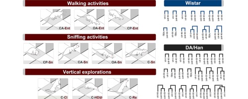
We have analyzed the temporal patterns of behaviour of male rats of the Wistar and DA/Han strains on the central platform of the elevated plus maze. The ethogram encompassed 10 behavioural elements. Durations, frequencies and latencies showed quantitative differences as to walking and sniffing activities. Wistar rats displayed significantly lower latency and significantly higher durations and frequencies of walking activities. DA/Han rats showed a significant increase of sniffing duration. In addition, DA/Han rats showed a significantly higher amount of time spent in the central platform. Multivariate T-pattern analysis revealed differences in the temporal organization of behaviour of the two rat strains. DA/Han rats showed (a) higher behavioural complexity and variability and (b) a significantly higher mean number of T-patterns than Wistar rats. Taken together, T-pattern analysis of behaviour in the centre of the elevated plus maze can noticeably improve the detection of subtle features of anxiety related behaviour. We suggest that T-pattern analysis could be used as sensitive tool to test the action of anxiolytic and anxiogenic manipulations.
Source:Journal of Neuroscience Methods, Volume 268
Author(s): M. Casarrubea, F. Faulisi, F. Caternicchia, A. Santangelo, G. Di Giovanni, A. Benigno, M.S. Magnusson, G. Crescimanno
We have analyzed the temporal patterns of behaviour of male rats of the Wistar and DA/Han strains on the central platform of the elevated plus maze. The ethogram encompassed 10 behavioural elements. Durations, frequencies and latencies showed quantitative differences as to walking and sniffing activities. Wistar rats displayed significantly lower latency and significantly higher durations and frequencies of walking activities. DA/Han rats showed a significant increase of sniffing duration. In addition, DA/Han rats showed a significantly higher amount of time spent in the central platform. Multivariate T-pattern analysis revealed differences in the temporal organization of behaviour of the two rat strains. DA/Han rats showed (a) higher behavioural complexity and variability and (b) a significantly higher mean number of T-patterns than Wistar rats. Taken together, T-pattern analysis of behaviour in the centre of the elevated plus maze can noticeably improve the detection of subtle features of anxiety related behaviour. We suggest that T-pattern analysis could be used as sensitive tool to test the action of anxiolytic and anxiogenic manipulations.
Graphical abstract
Tags: Neurology
from #Med Blogs by Alexandros G.Sfakianakis via Alexandros G.Sfakianakis on Inoreader http://ift.tt/25ZTp9N
via IFTTT
Δεν υπάρχουν σχόλια:
Δημοσίευση σχολίου