Σφακιανάκης Αλέξανδρος
ΩτοΡινοΛαρυγγολόγος
Αναπαύσεως 5 Άγιος Νικόλαος
Κρήτη 72100
00302841026182
00306932607174
alsfakia@gmail.com

Αρχειοθήκη ιστολογίου

! # Ola via Alexandros G.Sfakianakis on Inoreader

Η λίστα ιστολογίων μου

Κυριακή 24 Ιανουαρίου 2021

Expression of expandedFMR1-CGG repeats alters mitochondrial miRNAs and modulates mitochondrial functions and cell death in cellular model of FXTAS

xloma.fota13 shared this article with you from Inoreader

1-s2.0-S0891584921000551-fx1.jpg

Publication date: Available online 23 January 2021

Source: Free Radical Biology and Medicine

Author(s): Dhruv Gohel, Lakshmi Sripada, Paresh Prajapati, Fatema Currim, Milton Roy, Kritarth Singh, Anjali Shinde, Minal Mane, Darshan Kotadia, Flora Tassone, Nicolas Charlet-Berguerand, Rajesh Singh

View on the web

Human adventitial pericytes provide a unique source of anti-calcific cells for cardiac valve engineering: Role of microRNA-132-3p

xloma.fota13 shared this article with you from Inoreader

1-s2.0-S0891584921000460-fx1.jpg

Publication date: Available online 23 January 2021

Source: Free Radical Biology and Medicine

Author(s): Eva Jover, Marco Fagnano, William Cathery, Sadie Slater, Emanuela Pisanu, Yue Gu, Elisa Avolio, Domenico Bruno, Daniel Baz-Lopez, Ashton Faulkner, Michele Carrabba, Gianni Angelini, Paolo Madeddu

View on the web

Endothelium-dependent remote signaling in ischemia and reperfusion: alterations in the cardiometabolic continuum

xloma.fota13 shared this article with you from Inoreader

1-s2.0-S0891584921000575-fx1.jpg

Publication date: Available online 23 January 2021

Source: Free Radical Biology and Medicine

Author(s): Ralf Erkens, Matthias Totzeck, Amanda Brum, Dragos Duse, Hans Erik Bøtker, Tienush Rassaf, Malte Kelm

View on the web

Heme oxygenase-1(HO-1) regulates Golgi stress and attenuates endotoxin-induced acute lung injury through hypoxia inducible factor-1α (HIF-1α)/HO-1 signaling pathway

xloma.fota13 shared this article with you from Inoreader

1-s2.0-S0891584921000459-fx1.jpg

Publication date: Available online 23 January 2021

Source: Free Radical Biology and Medicine

Author(s): Xiangyun Li, Jianbo Yu, Lirong Gong, Yuan Zhang, Shuan Dong, Jia Shi, Cui Li, Yuting Li, Yanfang Zhang, Haibo Li

View on the web

Muscles in and around the ear as the source of “physiological noise” during auditory selective attention: a review and novel synthesis

xloma.fota13 shared this article with you from Inoreader

Abstract

The sensitivity of the auditory system is regulated via two major efferent pathways: the medial olivocochlear system that connects to the outer hair cells, and by the middle ear muscles – the tensor tympani and stapedius. The role of the former system in suppressing otoacoustic emissions has been extensively studied, but that of the complementary network has not. In studies of selective attention, decreases in otoacoustic emissions from contralateral stimulation have been ascribed to the medial olivocochlear system, but the acknowledged problem is that the results can be confounded by parallel muscle activity. Here, the potential role of the muscle system is examined through a wide but not exhaustive review of the selective attention literature, and the unifying hypothesis is made that the prominent "physiological noise" detected in such experiments, which is reduced during attention, is the sound produced by the muscles in proximity to the ear – including the middle ear m uscles. All muscles produce low‐frequency sound during contraction, but the implications for selective attention experiments – in which muscles near the ear are likely to be active – have not been adequately considered. This review and synthesis suggests that selective attention may reduce physiological noise in the ear canal by reducing the activity of muscles close to the ear. Indeed, such an experiment has already been done, but the significance of its findings have not been widely appreciated. Further sets of experiments are needed in this area.

View on the web

Aesthetic and therapeutic outcome of fat grafting for localized Scleroderma treatment: From basic study to clinical application

xloma.fota13 shared this article with you from Inoreader

Abstract

Background

Localized scleroderma (LoS) is a rare autoimmune disease characterized by skin fibrosis and subcutaneous tissue atrophy, resulting in aesthetic impairment on patients. Fat grafting has been used to treat LoS patients, achieving aesthetic and therapeutic improvement.

Aims

This article summarized the epidemiology and pathophysiology of LoS and the current progress and thorny questions of basic and clinical research on fat grafting treating LoS.

Methods

The literature of the last 20 years concerning fat grafting of treating LoS was reviewed.

Results

Fat grafting has been proved to produce aesthetic and therapeutic outcomes on LoS patients, including the improvement of soft tissue atrophy, skin fibrosis and pigmentation. Due to the inflammatory microenvironment of scleroderma, however, fat grafting still faces many difficulties, such as low fat retention. Novel fat grafting methods in order to supplement the deficiency of adipose‐derived stem cells and improve fat retention in LoS groups have been proposed whose effectiveness and feasibility is still needed further study.

Conclusion

Currently, fat grafting has been regarded as an effective treatment with a combination of aesthetic and therapeutic outcomes on LoS patients.

View on the web

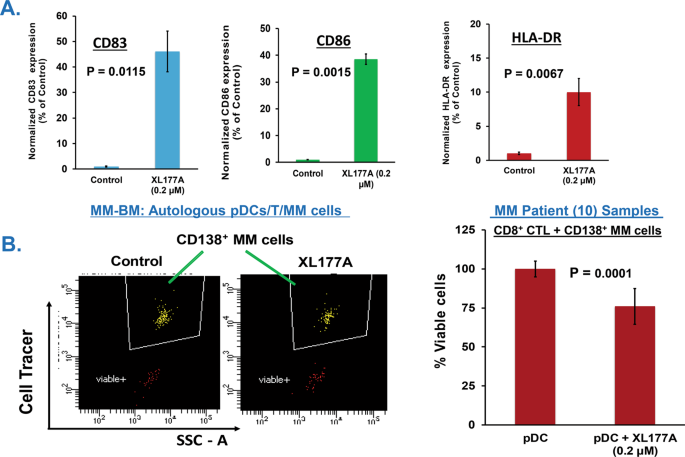
Targeting ubiquitin-specific protease-7 in plasmacytoid dendritic cells triggers anti-myeloma immunity

xloma.fota13 shared this article with you from Inoreader

41375_2021_1129_Fig1_HTML.png

View on the web

Periodontitis is a Factor Associated with Dyslipidemia

xloma.fota13 shared this article with you from Inoreader

Abstract

Objective

To investigate the association between the severity of periodontitis (exposure) and dyslipidemia (outcome).

Methods

This was a cross‐sectional study of users of public health services. Periodontitis was defined using the Center for Disease Prevention and Control and the American Academy of Periodontology criteria. Lipid evaluation used data on systemic biomarkers. Dyslipidemia diagnosis was based on the Guidelines of total cardiovascular risk of the World Health Organization. Weight, height, waist circumference, and blood pressure were measured, and socioeconomic‐demographic, lifestyle behavior factors, general and oral health conditions of the participants were collected. Hierarchical and logistic regression analyzes were used to determine the association between the exposures and the outcome. Odds Ratios, unadjusted and adjusted, and 95% confidence intervals were estimated.

Results

Of 1,011 individuals examined, 75.17% had dyslipidemia, and 84.17% had periodontitis, 0.2% with mild, 48.56% moderate and 35.41% severe disease. The association between periodontitis and dyslipidemia was maintained through hierarchical analysis and in the multiple regression modeling, showing that the occurrences of dyslipidemia in the group with periodontitis, and its moderate and severe levels, were, respectively, 14%, 30%, and 16% higher compared with those without periodontitis.

Conclusions

The results showed a positive association between moderate and severe periodontitis and dyslipidemia.

View on the web

Effects of ibuprofen administration timing on oral surgery pain: A randomized clinical trial

xloma.fota13 shared this article with you from Inoreader

Abstract

Objective

This study aimed to compare the analgesic effect of ibuprofen 400 mg given 30 minutes before or immediately after third molars surgery under local anaesthesia.

Materials and Methods

The single‐center, randomized, split‐mouth, triple‐blind, clinical trial involved 38 outpatients, for a total of 76 bilateral symmetrical fully bone impacted mandibular third molars. Each patient was undergone to separate surgical sessions for the right and left side, and ibuprofen was randomly administered 30 minutes before or immediately after the intervention. Study participants recorded pain intensity using Numerical Rating Scale‐11, the timing of rescue therapy intake, and overall tablets consumption over three days.

Results

The overall pain intensity score was lower in the group receiving ibuprofen immediately after (3.13 ± 2.46) than before (3.58 ± 2.40) surgery, with statistically significant differences only on the second and third day. The mean time to the first using rescue therapy was longer in the postoperative (598.33 ± 422.62 minutes) than in the preoperative (406.25 ± 149.79 minutes) analgesic treatment group (p=0.123). The number of supplemented ibuprofen tablets did not differ (p=0.530) between both groups.

Conclusions

Within the limits of the present study, ibuprofen administration immediately after surgery seemed to be more effective than preoperative administration.

View on the web

The systemic inflammation response index predicts the survival of patients with clinical T1‐2N0 oral squamous cell carcinoma

xloma.fota13 shared this article with you from Inoreader

Abstract

Objective

The systemic inflammation response index (SIRI) is an independent prognostic factor for many malignant tumors. However, the value of this factor in patients with clinical T1‐2N0 (cT1‐2N0) oral squamous cell carcinoma (OSCC) is still unclear.

Methods

We calculated SIRI of 235 cT1‐2N0 OSCC patients from 2013 to 2017. Multivariate cox regression analysis was applied to verify the prognostic significance of SIRI. Kaplan‐Meier curves were plotted to analyze the overall survival (OS) and disease‐specific survival (DSS) for cT1‐2N0 OSCC patients.

Results

According to the optimal cutoff point of SIRI, we divided cT1‐2N0 OSCC patients into high SIRI group (SIRI ≥ 1.3) and low SIRI group (SIRI < 1.3). SIRI was an independent prognostic indicator for OS (HR = 2.87; 95% CI = 1.35‐6.10; P = 0.006) and DSS (HR = 2.17; 95% CI = 1.10‐4.27; P = 0.025). High SIRI had a significantly poorer OS (P = 0.001) and DSS (P = 0.007) in survival analysis than the low SIRI. Moreover, the prognostic value of SIRI was significantly stronger than neutrophil‐to‐lymphocyte ratio (NLR) and monocyte‐to‐lymphocyte ratio (MLR).

Conclusions

Preoperative SIRI can be regarded as a meaningful indicator for poor survival of cT1‐2N0 OSCC patients, and it is a promising tool to formulate the best individualized treatment for high‐risk patients.

View on the web

Age‐related dental phenotypes and tooth characteristics of FAM83H‐associated hypocalcified amelogenesis imperfect

xloma.fota13 shared this article with you from Inoreader

Abstract

Objectives

Autosomal‐dominant hypocalcified amelogenesis imperfecta (ADHCAI) shows phenotypic heterogeneity. Our aim was to characterise the ADHCAI phenotypes, tooth properties, and genotypes.

Methods

Three unrelated ADHCAI probands and seven additional affected members of the three families were recruited. Mutations were identified by exome and Sanger sequencing, and haplotypes by SNP array. Tooth colour, roughness, density, nanohardness, minerals, and ultrastructure were investigated.

Results

Ten participants were heterozygous for the FAM83H mutation c.1387C>T (p.Gln463*). All shared a 3.43 Mbp region on chromosome 8q24.3 encompassing the FAM83H variant, indicating a common ancestry. The c.1387C>T was estimated to be 23.8 generations or 600 years. The FAM83H enamel had higher roughness and lower lightness, density, nanohardness, and calcium and phosphorus levels than controls. Blunted enamel rods, wide interrod spaces, and disorganized dentinoenamel junctions were observed. Evaluating the patients with the same mutation and reviewing others with different mutations in FAM83H revealed that the FAM83H heterogeneous phenotypes are age‐influenced. Tooth colour and surface texture change with aging.

Conclusions

FAM83H enamel demonstrated decreased lightness, density, hardness, calcium, phosphorus, and defective ultrastructure. We have identified that the phenotypic variation in FAM83H‐associated ADHCAI is age‐related. Awareness of the correlation between age and clinical features of FAM83H‐ADHCAI can help dentists make an accurate diagnosis.

View on the web

Efficacy and safety of sorafenib plus vitamin K treatment for hepatocellular carcinoma: A phase II, randomized study

xloma.fota13 shared this article with you from Inoreader
Efficacy and safety of sorafenib plus vitamin K treatment for hepatocellular carcinoma: A phase II, randomized study

Vitamin K dosing during sorafenib treatment against HCC showed higher ORR and prolonged PFS significantly. The vitamin K dosing might enhance the antitumor action of sorafenib by suppressing des‐γ‐carboxy prothrombin, which is a factor that promotes angiogenesis and tumor growth.


Abstract

The previous retrospective study suggested that dosing vitamin K may enhance the anticancer action of sorafenib against hepatocellular carcinoma. To confirm it, we performed a phase II, randomized, open‐label study. Patients with hepatocellular carcinoma were randomly assigned to receive sorafenib + vitamin K2 (menatetrenone, 45 mg daily, orally) or sorafenib only. Between 1 May 2012 and 1 May 2016, 68 patients were screened. Forty‐four eligible patients were assigned at a 1:1 ratio to each cohort. The objective response rate in the vitamin K‐dosed group was significantly higher than that in the sorafenib only group (27.3% vs 4.5%, respectively; p = 0.039). The median time of progression‐free survival was significantly extended in the vitamin K‐dosed group compared with the sorafenib only group (4.9 months vs 2.7 months, respectively; hazard ratio (HR), 0.44; 95% confidence interval (CI): 0.21–0.89; p = 0.018). Although there was no significant difference between the two groups in the median time of overall survival, patients in the vitamin K‐dosed group with a complete response or partial response achieved a significantly extended median time of overall survival compared with the other patients in the vitamin K‐dosed group or the patients in the sorafenib only group (26.1 months vs 9.0 months; HR, 0.34; 95% CI: 0.11–0.95; p = 0.046 or 11.5 months; HR, 0.16; 95% CI: 0.034–0.70; p = 0.006, respectively). Dosing vitamin K could augment the anticancer action of sorafenib against HCC.

View on the web

Αρχειοθήκη ιστολογίου