Σφακιανάκης Αλέξανδρος
ΩτοΡινοΛαρυγγολόγος
Αναπαύσεως 5 Άγιος Νικόλαος
Κρήτη 72100
00302841026182
00306932607174
alsfakia@gmail.com

Αρχειοθήκη ιστολογίου

! # Ola via Alexandros G.Sfakianakis on Inoreader

Η λίστα ιστολογίων μου

Δευτέρα 23 Μαΐου 2022

Comparison between the radial forearm and groin soft tissue free flaps for reconstruction in patients with oral cavity cancer: a quality of life analysis

alexandrossfakianakis shared this article with you from Inoreader
The purpose of this study was to compare the effects of the radial forearm free flap (RFFF) and groin soft tissue free flap (GSFF) on the quality of life (QoL) of patients undergoing reconstructive surgery after resection for oral cancer. A retrospective analysis of 48 patients was performed. The Vancouver Scar Scale (VSS), University of Washington Quality of Life (UW-QOL) questionnaire, and 14-item Oral Health Impact Profile (OHIP-14) questionnaire were used to evaluate the donor site scars and QoL of the patients. (Source: International Journal of Oral and Maxillofacial Surgery)
View on Web

Promoter hypermethylation of GALR1 acts as an early epigenetic susceptibility event in colorectal carcinogenesis

alexandrossfakianakis shared this article with you from Inoreader
View on Web

Κυριακή 22 Μαΐου 2022

Activation of the mitogen‐activated protein kinase–extracellular signal‐regulated kinase pathway in childhood B‐cell acute lymphoblastic leukemia

alexandrossfakianakis shared this article with you from Inoreader

Abstract

RAS mutations are frequently observed in childhood B-cell acute lymphoblastic leukemia (B-ALL) and previous studies have yielded conflicting results as to whether they are associated with a poor outcome. We and others have demonstrated that the mitogen-activated protein kinase–extracellular signal-regulated kinase (MAPK) pathway can be activated through epigenetic mechanisms in the absence of RAS pathway mutations. Herein, we examined whether MAPK activation, as determined by measuring phosphorylated extracellular signal-regulated kinase (pERK) levels in 80 diagnostic patient samples using phosphoflow cytometry, could be used as a prognostic biomarker for pediatric B-ALL. The mean fluorescence intensity of pERK (MFI) was measured at baseline and after exogenous stimulation with or without pretreatment with the mitogen-activated protein kinase kinase (MEK) inhibitor trametinib. Activation levels (MFI stimulated/MFI baseline) ranged from 0.76 to 4.40 (median =&n bsp;1.26), and inhibition indexes (MFI stimulated/MFI trametinib stimulated) ranged from 0.439 to 5.640 (median = 1.30), with no significant difference between patients with wildtype versus mutant RAS for either. Logistic regression demonstrated that neither MAPK activation levels nor RAS mutation status at diagnosis alone or in combination was prognostic of outcome. However, 35% of RAS wildtype samples showed MAPK inhibition indexes greater than the median, thus raising the possibility that therapeutic strategies to inhibit MAPK activation may not be restricted to patients whose blasts display Ras pathway defects.

View on Web

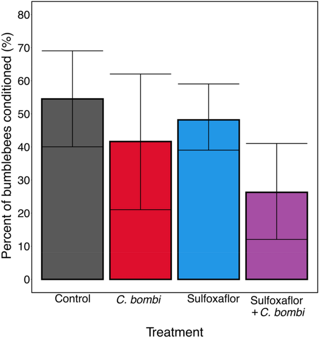
No effect of dual exposure to sulfoxaflor and a trypanosome parasite on bumblebee olfactory learning

alexandrossfakianakis shared this article with you from Inoreader

41598_2022_12714_Fig1_HTML.png

View on Web

Application of an Environmental Monitoring to Assess the Practices and Control the Risk of Occupational Exposure to Cyclophosphamide in Two Sites of a French Comprehensive Cancer Center

alexandrossfakianakis shared this article with you from Inoreader
Abstract
Objectives
The risk of chronic exposure to antineoplastic agents in hospitals, mainly by skin contact with contaminated surfaces, is well established. The aim of this study was to assess indirectly the risk of occupational exposure to antineoplastics drugs at two hospitals by using an environmental monitoring, and to suggest ways of improving the exposure to healthcare workers.
Methods
An observational study of care practices on both sites was carried out. A wipe sampling campaign was then designed to study environmental contamination throughout the chemotherapy process: receipt, storage, compounding, transport, administration, and elimination areas. Samples were analyzed by a validated LC-MS/MS method allowing trace quantification of cyclophosphamide. A guidance 'safe value' of 0.10 ng/cm2 was considered.
Results
A total of 293 samples were analyzed, of which 58% were found to be positive. In the compou nding units, the drug vials were contaminated before [range = (non-quantifiable [NQ]-0.71) ng/cm2] and after cleaning procedure [(NQ-0.62) ng/cm2], particularly when the flip-off lid was removed during cleaning. The contamination found on manual preparations was operator-dependent: [non-detectable (ND)-3.51] ng/cm2 on infusion bag surfaces; (780.61–24 698.98) ng/cm2 on medication ports. In the case of automated preparations, the average contamination was higher on infusion bag surfaces [(2.43–36.86) ng/cm2] and lower on medication ports [(0.43–7.65) ng/cm2] than manual preparations. Contamination of the analytical control area was also highlighted. In the daily care unit, the contamination was located near the infusion area (armchairs, infusion stands, floor, and patient toilets), and varied somewhat between the two sites, especially on the floor with (0.46–27.32) compared to (ND-0.18) ng/cm2. We di d not detect contamination on the transport boxes, on the door handles or in the disposal areas.
Conclusions
The variability of contamination observed between the two sites can be explained in part by the difference in routine practices, especially training of the staff, and cleaning procedures. Findings were communicated to healthcare workers, and news interventions were implemented based on wipe sampling results. This study demonstrated a method for routine environmental monitoring and worker education as a strategy to reduce occupational exposure.

View on Web

Diagnostic potential of extracellular vesicles in meningioma patients

alexandrossfakianakis shared this article with you from Inoreader
Abstract
Background
Extracellular vesicles (EVs) play an important role in cell-cell communication, and tumor-derived EVs circulating in patient blood can serve as biomarkers. Here, we investigated the potential role of plasma EVs in meningioma patients for tumor detection and determined whether EVs secreted by meningioma cells reflect epigenetic, genomic and proteomic alterations of original tumors.
Methods
EV concentrations were quantified in patient plasma (n = 46). Short-term meningioma cultures were established (n = 26) and secreted EVs were isolated. Methylation and copy number profiling was performed using 850k arrays, and mutations were identified by targeted gene panel sequencing. Differential quantitative mass spectrometry was employed for proteomic analysis.
Results
Levels of circulating EVs were elevated in meningioma patients compared to healthy individuals, and the plasma EV concentration correlated with malignanc y grade and extent of peritumoral edema. Postoperatively, EV counts dropped to normal levels, and the magnitude of the postoperative decrease was associated with extent of tumor resection. Methylation profiling of EV-DNA allowed correct tumor classification as meningioma in all investigated cases, and accurate methylation subclass assignment in almost all cases. Copy number variations present in tumors, as well as tumor-specific mutations were faithfully reflected in meningioma EV-DNA. Proteomic EV profiling did not permit original tumor identification but revealed tumor-associated proteins that could potentially be utilized to enrich meningioma EVs from biofluids.
Conclusions
Elevated EV levels in meningioma patient plasma could aid in tumor diagnosis and assessment of treatment response. Meningioma EV-DNA mirrors genetic and epigenetic tumor alterations and facilitates molecular tumor classification.
View on Web

Characteristics of Children ≤36 Months of Age with Diffuse Intrinsic Pontine Glioma (DIPG): A Report from the International DIPG Registry

alexandrossfakianakis shared this article with you from Inoreader
ABSTRACT:
Background
Children ≤36 months with Diffuse Intrinsic Pontine Glioma (DIPG) have increased long-term survival (LTS, overall survival (OS) ≥24 months). Understanding distinguishing characteristics in this population is critical to improving outcomes.
Methods
Patients ≤36 months at diagnosis enrolled on the International DIPG Registry (IDIPGR) with central imaging confirmation were included. Presentation, clinical course, imaging, pathology and molecular findings were analyzed.
Results
Among 1183 patients in IDIPGR, 40 were eligible (median age: 29 months). Median OS was 15 months. Twelve patients (30%) were LTS, 3 (7.5%) very long-term survivors ≥ 5 years. Among 8 untreated patients, median OS was 2 months. Patients enrolled in the registry but excluded from our study by central radiology review or tissue diagnosis had median OS of 7 months. All but 1 LTS received radiation. Among 32 treated patients, 1-, 2-, 3 -, and 5-year OS rates were 68.8%, 31.2%, 15.6% and 12.5%, respectively. LTS had longer duration of presenting symptoms (p=0.018). No imaging features were predictive of outcome. Tissue and genomic data were available in 18 (45%) and 10 patients, respectively. Among 9 with known H3K27M status, 6 had a mutation.
Conclusions
Children ≤36 months demonstrated significantly more LTS, with an improved median OS of 15 months; 92% of LTS received radiation. Median OS in untreated children was 2 months, compared to 17 months for treated children. LTS had longer duration of symptoms. Excluded patients demonstrated a lower OS, contradicting the hypothesis that children ≤36 months with DIPG show improved outcomes due to misdiagnosis.
View on Web

Αρχειοθήκη ιστολογίου