Σφακιανάκης Αλέξανδρος
ΩτοΡινοΛαρυγγολόγος
Αναπαύσεως 5 Άγιος Νικόλαος
Κρήτη 72100
00302841026182
00306932607174
alsfakia@gmail.com

Αρχειοθήκη ιστολογίου

! # Ola via Alexandros G.Sfakianakis on Inoreader

Η λίστα ιστολογίων μου

Τρίτη 19 Ιουλίου 2022

Differential avidity determination of IgG directed towards the receptor‐binding domain (RBD) of SARS‐CoV‐2 wildtype and its variants in one assay: Rational tool for the assessment of protective immunity.

alexandrossfakianakis shared this article with you from Inoreader

Abstract

The avidity (binding strength) of IgG directed towards the receptor-binding domain (RBD) of spike protein has been recognized as a central marker in SARS-CoV-2 serology. It seems to be linked to increased infection-neutralization potential and therefore might indicate protective immunity. Using a prototype line assay based on the established recomLine SARS-CoV-2 assay, supplemented with RBD of the delta and the omicron variant, differential avidity determination of IgG directed towards RBD of wild-type (WT) SARS-CoV-2 and distinct variants was possible within one assay. Our data confirm that natural SARS-CoV-2 infection or one vaccination step lead to low avidity IgG, whereas further vaccination steps gradually increase avidity to high values. High avidity is not reached by infection alone. After infection with WT SARS-CoV-2 or vaccination based on mRNA WT, the avidity of cross-reacting IgG directed towards RBD of the delta variant only showed marginal diffe rences compared to IgG directed towards RBD WT. In contrast, the avidity of IgG cross-reacting with RBD of the omicron variant was always much lower than for IgG RBD WT, except after the third vaccination step. Therefore, parallel avidity testing of RBD WT and omicron seems to be mandatory for a significant assessment of protective immunity towards SARS-CoV-2.

This article is protected by copyright. All rights reserved.

View on Web

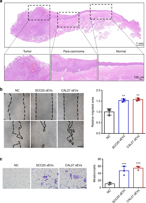
Cancer cells corrupt normal epithelial cells through miR-let-7c-rich small extracellular vesicle-mediated downregulation of p53/PTEN

alexandrossfakianakis shared this article with you from Inoreader

41368_2022_192_Fig1_HTML.png

International Journal of Oral Science, Published online: 19 July 2022; doi:10.1038/s41368-022-00192-2

Cancer cells corrupt normal epithelial cells through miR-let-7c-rich small extracellular vesicle-mediated downregulation of p53/PTEN
View on Web

New insight on the role of Helicobacter pylori cagA in the expression of cell surface antigens with important biological functions in gastric carcinogenesis

alexandrossfakianakis shared this article with you from Inoreader

Abstract

Background

Expression of cluster of differentiation (CD) antigens changes according to disease status and inflammation. Profiles of CD antigens expression in gastric cancer patients are different based on the status of H. pylori infection.

Aims

We conducted this study to profile CD antigen markers in gastric adenocarcinoma cells (AGS cell line) infected with distinct cytotoxin-associated gene A (cagA) genotypes of H. pylori clinical isolates.

Methods

The AGS cells were infected with H. pylori isolates with different cagA genotypes, and CD antigens expression was determined using DotScan™ antibody microarray. Formation of "hummingbird" phenotype was determined, and the percentage was calculated.

Results

H. pylori strains harboring cagA upregulated the expression of CD antigen involved in cancer stem cell formation (CD55), but downregulated CD antigens involved in immune regulation (CD40 and CD186) and cell adhesion (CD44). CD54 (neutrophil adhesion) and CD71 (iron transfer) were highly downregulated in the gastric cells infected with Western cagA isolates compared with East Asian isolates. CD antigen expression was different in the cells infected with H. pylori harboring different CagA EPIYA (Glu-Pro-Ile-Tyr-Ala) numbers, in which higher repression of CD54 and CD15 (Lewis x antigen) were observed in the isolate with the highest number of EPIYA motif. Furthermore, higher downregulation of CD15 was observed in the infected gastric cells with high percentage of "hummingbird" phenotype than that of low percentage of "hummingbird" phenotype.

Conclusion

Our study demonstrated the critical roles of CD antigens in the CagA pathogenesis and should be investigated further.

View on Web

Comparative study of DFAT cell and ADSC sheets for periodontal tissue regeneration:in vivo and in vitro evidence

alexandrossfakianakis shared this article with you from Inoreader

Abstract

Aim

To compare the efficacy of adipocyte-derived dedifferentiated fat (DFAT) cell and adipose-derived stromal cell (ADSC) sheets for regenerative treatment of intrabony periodontal defects.

Material and Methods

DFAT cells were obtained using the ceiling culture method and were compared with ADSCs using Cell Counting Kit-8 (CCK8), colony formation assay, surface antigen identification, and multilineage differentiation assays. DFAT and ADSC sheets were prepared in cell sheet culture medium. The biological characteristics of DFAT cell and ADSC sheets were compared using haematoxylin and eosin staining, quantitative reverse transcription PCR, and immunofluorescence staining. Micro-computed tomography and histological staining were used to compare the effects of the two cell sheets on the repair of periodontal intrabony defects in rats.

Results

DFAT cells and ADSCs demonstrated mesenchymal stem cell characteristics. Both cell type were CD29-, CD90-, and CD146-positive and CD31-, CD34-, and CD45-negative. DFAT cells and ADSCs exhibited similar osteogenic and adipogenic differentiation capabilities, and colony-formation ability. DFAT cells displayed stronger proliferation capabilities compared to ADSCs. Compared with the ADSC sheets, DFAT cell sheets exhibited a higher expression of periodontal-related genes and proteins and greater ability to regenerate periodontal tissue.

Conclusion

Our findings suggest that DFAT cell sheets are an ideal seed cell source and form of cell delivery for periodontal intrabony defects.

View on Web

The accuracies of three intraoral scanners with regards to shade determination: An in vitro study

alexandrossfakianakis shared this article with you from Inoreader

Abstract

Purpose

: To compare the accuracy of three intraoral scanners for shade determination function in vitro, and to preliminarily investigate the shade-matching characteristics of the three intraoral scanners.

Materials & Methods

The shade of the middle third region of each shade tab on the Vita Classical A1-D4 shade guide (VC) was measured by a spectrophotometer (Vita Easyshade V, VE) and three intraoral scanners, including CEREC Omnicam (OM), 3Shape TRIOS 3 (T3), and TRIOS 4 (T4). A conversion table between VC values and CIELAB values was established from the database of VE to analyze the trueness. The reproducibility of the instruments was then compared by repeating the measurements five times.

Results

: The mean color difference for each instrument was highest in the OM, followed by the T4, and lowest in the T3 and VE, repectively. The L* and a* value for OM, and the b* value for T4, were significantly different from those for VE (p <0.05). The reproducibility of the instrument was highest in the VE (Fleiss' kappa: 0.95), followed by the T3 (Fleiss' kappa: 0.89), T4 (Fleiss' kappa: 0.87), and OM (Fleiss' kappa: 0.78).

Conclusions

: Of the three intraoral scanners, the trueness was best on the T3. The reproducibility of all the instruments was excellent.

This article is protected by copyright. All rights reserved

View on Web

TREM2‐induced activation of microglia contributes to synaptic integrity in cognitively intact aged individuals with Alzheimer's neuropathology

alexandrossfakianakis shared this article with you from Inoreader
TREM2-induced activation of microglia contributes to synaptic integrity in cognitively intact aged individuals with Alzheimer's neuropathology

Efficient TREM2-phagocytic microglia underlie synaptic resilience in NDAN, removing damaged synpases, contributing to synaptic integrity and protecting from memory deficits.


Abstract

The existence of individuals who remain cognitively intact despite presenting histopathological signs of Alzheimer's disease (AD), here referred to as "Nondemented with AD neuropathology" (NDAN), suggests that some mechanisms are triggered to resist cognitive impairment. Exposed phosphatidylserine (ePS) represents a neuronal "eat-me" signal involved in microglial-mediated phagocytosis of damaged synapses. A possible mediator of this process is TREM2, a microglial surface receptor activated by ligands including PS. Based on TREM2 role in the scavenging function of microglia, we hypothesize that an efficient microglial phagocytosis of damaged synapses underlies synaptic resilience in NDAN, thus protecting from memory deficits. Using immunofluorescence microscopy, we performed a comparative study of human post-mortem frontal cortices of aged-matched, AD and NDAN individuals. We studied the distribution of activated microglia (IBA1, IBA1+/CD68+ cells) and phagocytic microglia-related proteins (TREM2, DAP12), demonstrating higher microglial activation and TREM2 expression in NDAN versus AD. A study of the preservation of synapses around plaques, assessed using MAP2 and βIII tubulin as dendritic and axonal markers, respectively, and PSD95 as a postsynaptic marker, revealed preserved axonal/dendritic structure around plaques in NDAN versus AD. Moreover, high levels of PSD95 around NDAN plaques and the colocalization of PSD95 with CD68 indicated a prompt removal of damaged synapses by phagocytic microglia. Furthermore, Annexin V assay on aged-matched, AD and NDAN individuals synaptosomes revealed increased levels of ePS in NDAN, confirming damaged synapses engulfment. Our results suggest a higher efficiency of TREM2-induced phagocytic microglia in removing damaged synapses, underlying synaptic resilience in NDAN individuals.

View on Web

A higher tumor volume and undernutrition at diagnosis adversely affect the survival of children with Wilms tumor: A study of 200 patients

alexandrossfakianakis shared this article with you from Inoreader

Abstract

Background

Distinct prognostic factors for Wilms tumor (WT) in low- and middle-income countries need identification.

Methods

Retrospective study of patients with WT managed by the International Society of Pediatric Oncology (SIOP) approach for over 11 years (2005–2016) at a single center in Chandigarh, India.

Results

The study included 200 patients (median age: 33.5 months). The tumor stage (SIOP) distribution included stage I (30%), II (36%), III (14%), IV (17%), and V (3%). The histology-risk groups were low (8%), intermediate (84%), and high risk (9%). At diagnosis, 68 out of 190 (36%) patients were underweight. The median tumor volume at diagnosis was 481 ml (interquartile ratio [IQR]: 306.9, 686.8, n = 146). Following neoadjuvant chemotherapy, it reduced to 110 ml (IQR: 151.2, 222, n = 77). Treatment was abandoned in 20.5% of the patients. Treatment-related mortality occurred in 13 of 179 (7.2%) patients. Relapse occurred in 26 of 158 (16.5%) patients. The 3-year overall survival (OS) and event-free survival (EFS) of patients who completed therapy were 78.3 and 72%, respectively. The stage (p = .013) and histology (p = .023) influenced OS. A lower OS in stage II (75.4%) versus stage III disease (83.7%) suggested understaging. Patients with a higher tumor volume at diagnosis (p = .005; odds ratio [OR]: 0.99; 95% confidence interval [CI]: 0.99–1.00) or a lower weight-for-age z-score (p = .002; OR: 1.68; 95% CI: 1.21–2.33) had an increased risk of death or relapse.

Conclusions

The 3-year OS and EFS of children who completed therapy were 78.3 and 72%, respectively. A higher tumor volume and lower weight-for-age z-score at diagnosis were identified as distinct adverse prognostic factors. A likely suboptimal lymph node assessment (intraoperative and histopathology) contributed to the understaging of stage III to II disease and reduced survival.

View on Web

Αρχειοθήκη ιστολογίου